MIROTECAMIROTECA
Real X-rays and CT images, each with a question, what it shows and a link to its classification chart. Try to classify the image before opening the answer.
The images come from Wikimedia Commons under free licenses (CC BY-SA), with the author and license under each one. They are not owned by MIROTECA. Where an image does not allow two types to be told apart, the question does not ask for it.
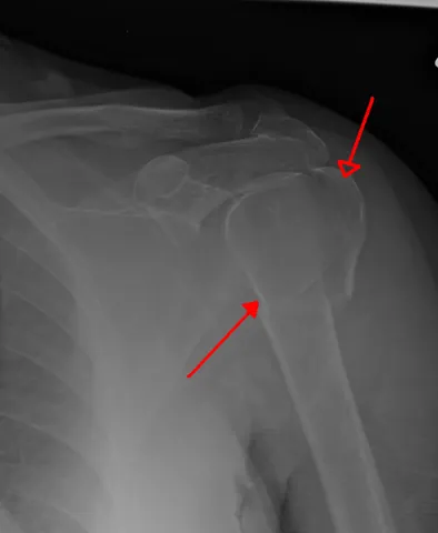 Three-part proximal humerus fracture on X-ray (Neer classification)
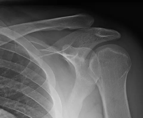 Grade III acromioclavicular joint separation on X-ray (Rockwood)
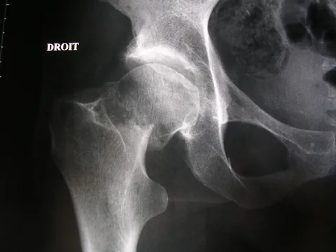 Displaced femoral neck fracture on X-ray (Garden classification)
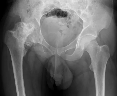 Collapsed femoral head avascular necrosis on X-ray (Ficat and Arlet)
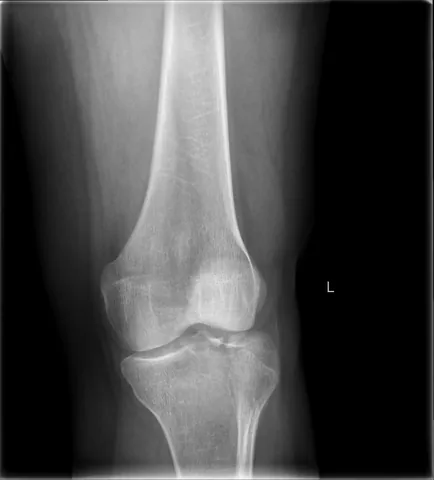 Lateral tibial plateau fracture with depression on X-ray (Schatzker)
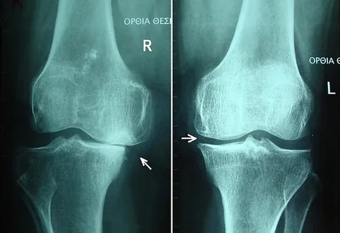 Knee osteoarthritis on a weight-bearing X-ray (Kellgren-Lawrence)
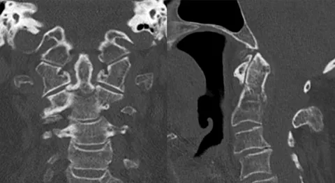 Type II odontoid fracture on CT (Anderson-D'Alonzo)
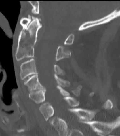 Type III odontoid fracture on CT (Anderson-D'Alonzo)
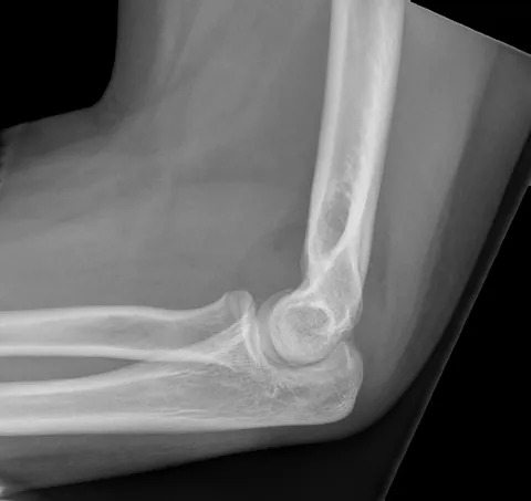
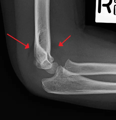 Sail sign in a child elbow: nondisplaced supracondylar fracture (Gartland I)
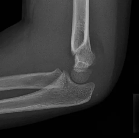 Displaced supracondylar fracture in a child: the anterior humeral line