
Clinical classifications are easier to remember as a picture than as a list. Each diagram below condenses one of them into a single image, with the cut-offs and the detail that usually decides the answer, followed by a short explanation of how to read it, the most common mistakes and how it tends to appear in exam questions.
The diagrams are free to download and share for study and teaching, with attribution. They are written and reviewed by the MIROTECA medical team, a group of doctors in training and in practice. MIROTECA started in Spanish, as a tool to practice clinical scales for the Spanish residency exam (MIR); this English section is new and will grow.
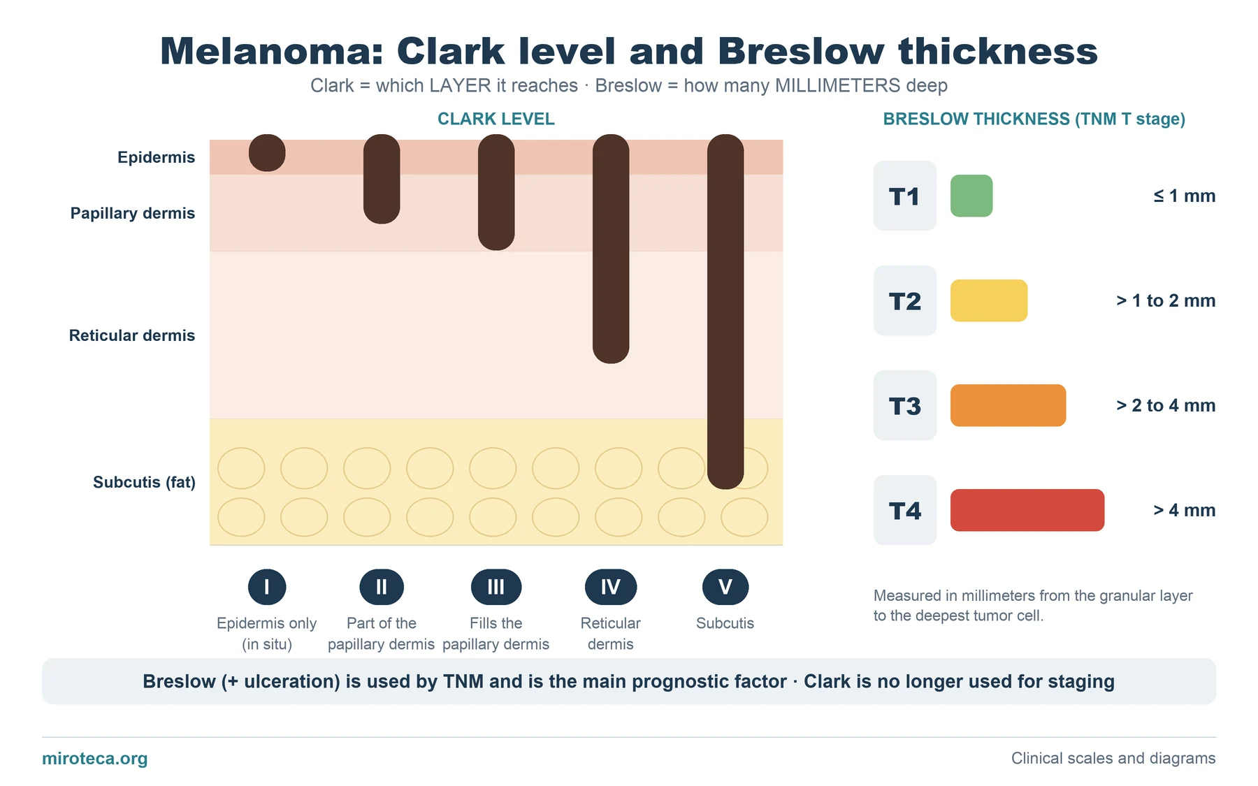 Breslow thickness and Clark level: melanoma diagram
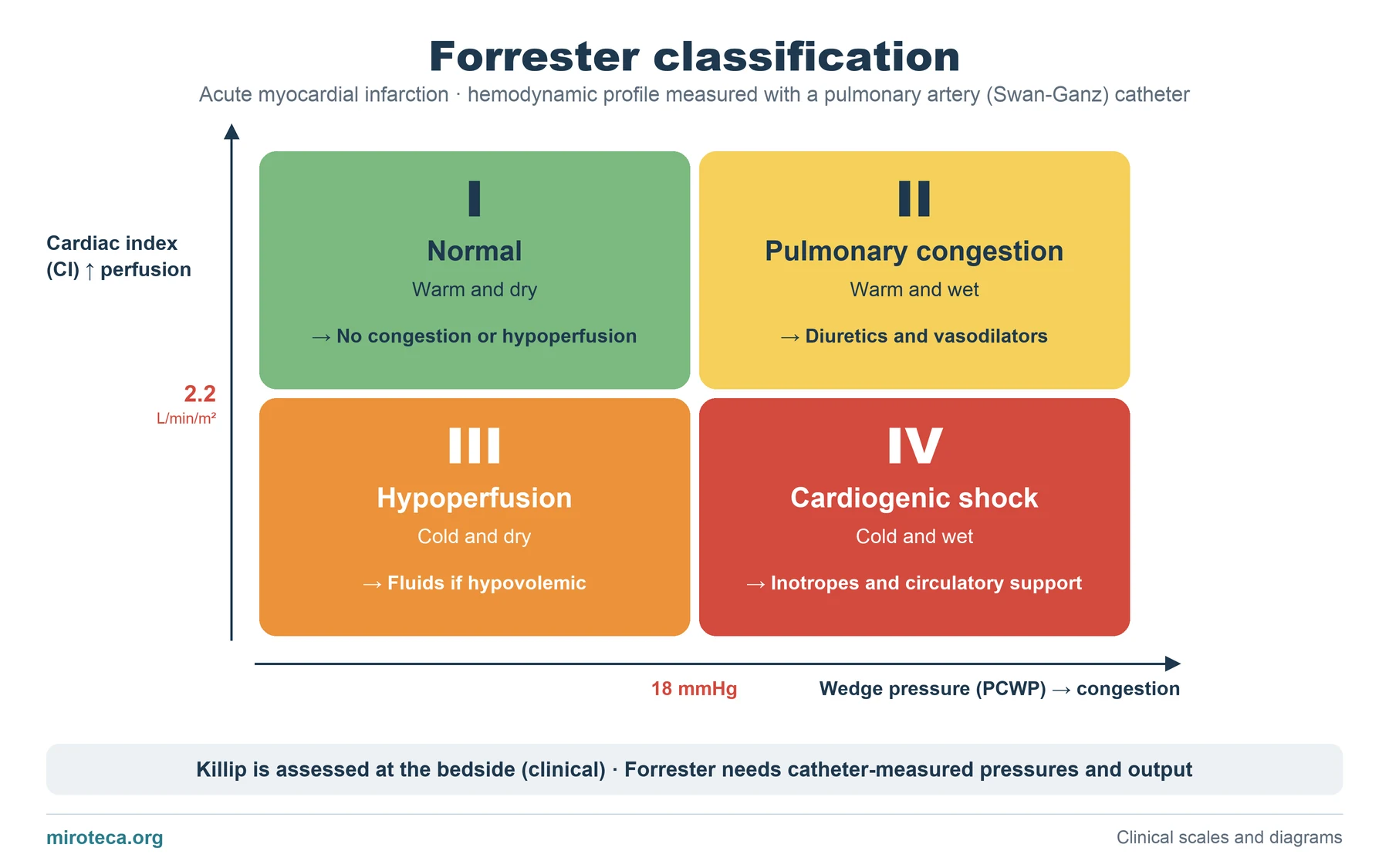 Forrester classification: the 4 hemodynamic subsets
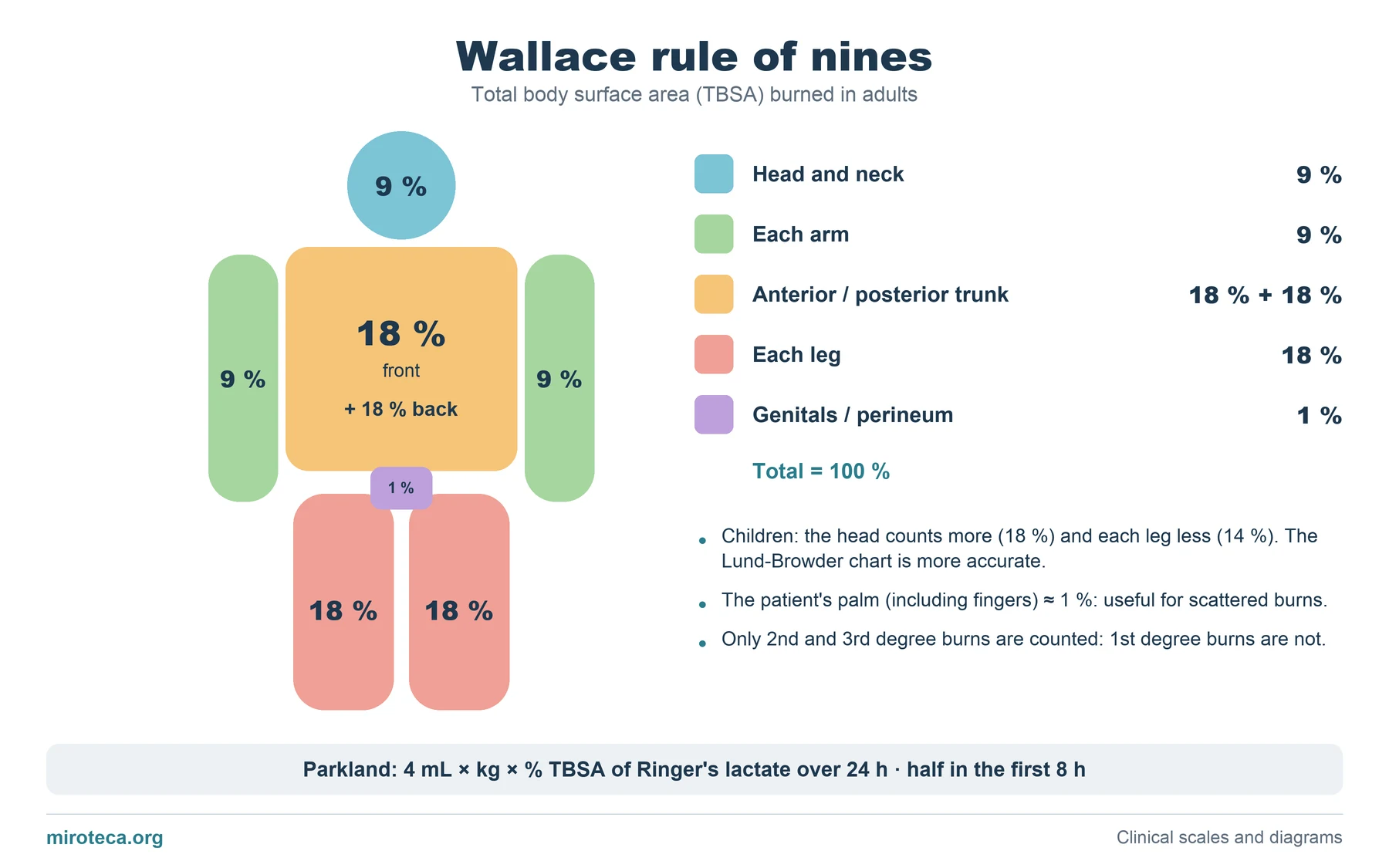 Rule of nines: burned body surface area diagram
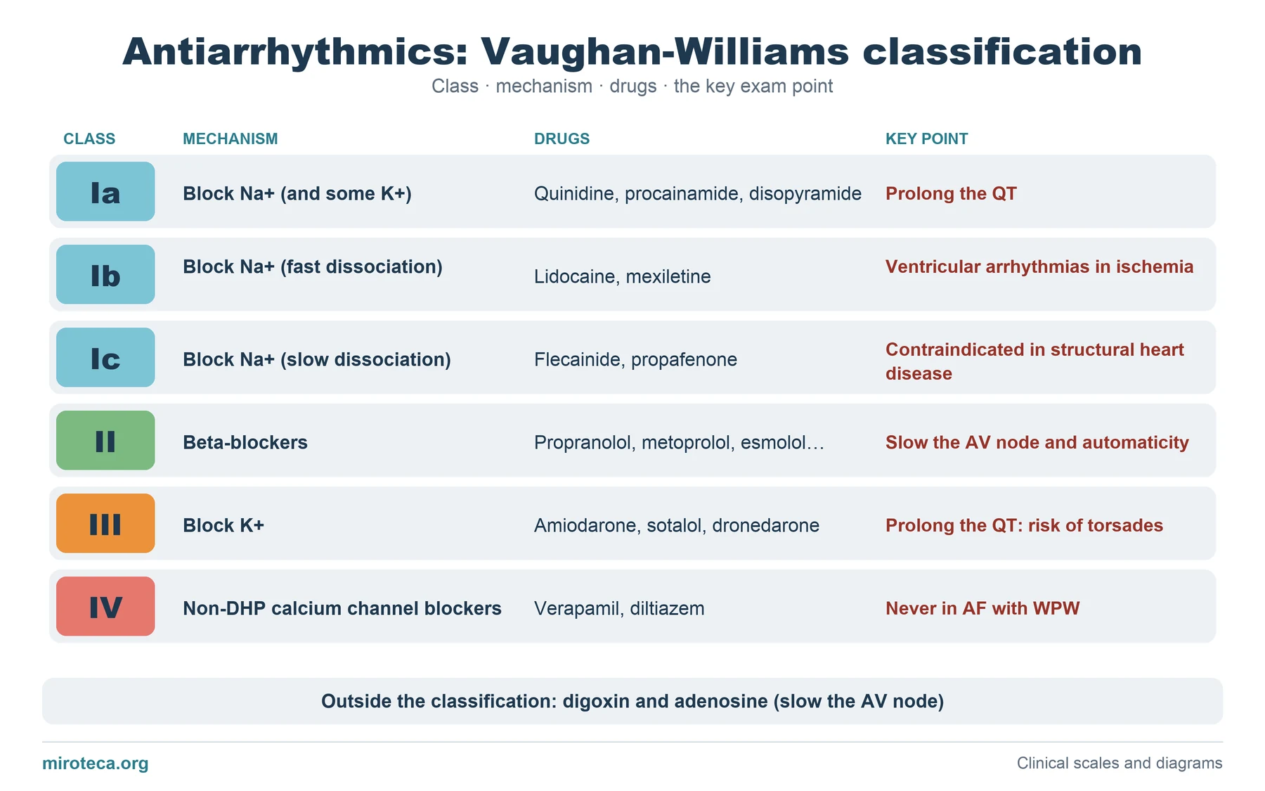 Vaughan-Williams classification of antiarrhythmic drugs
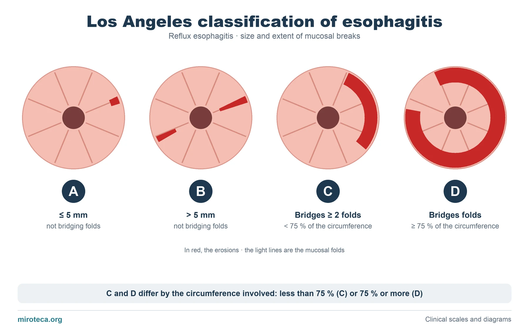 Los Angeles classification of esophagitis: grades A to D
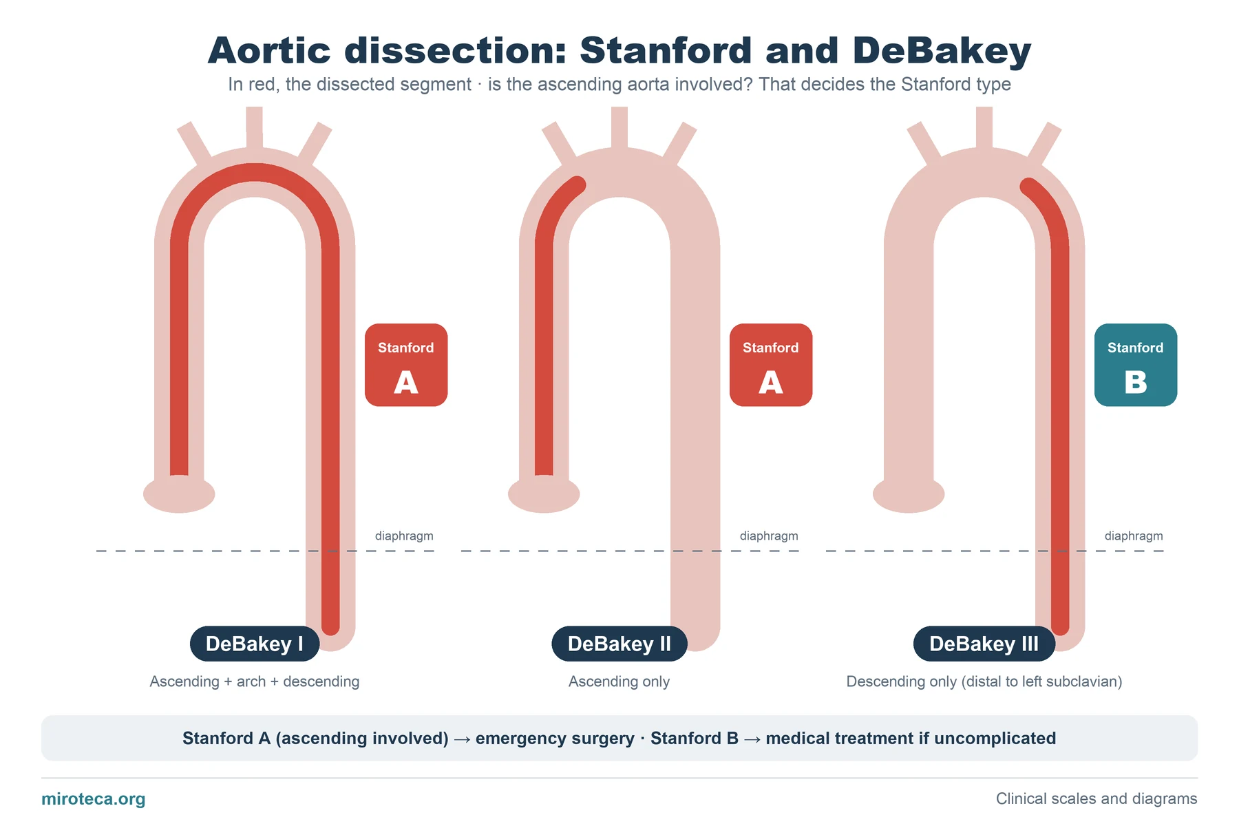 Stanford and DeBakey classification of aortic dissection
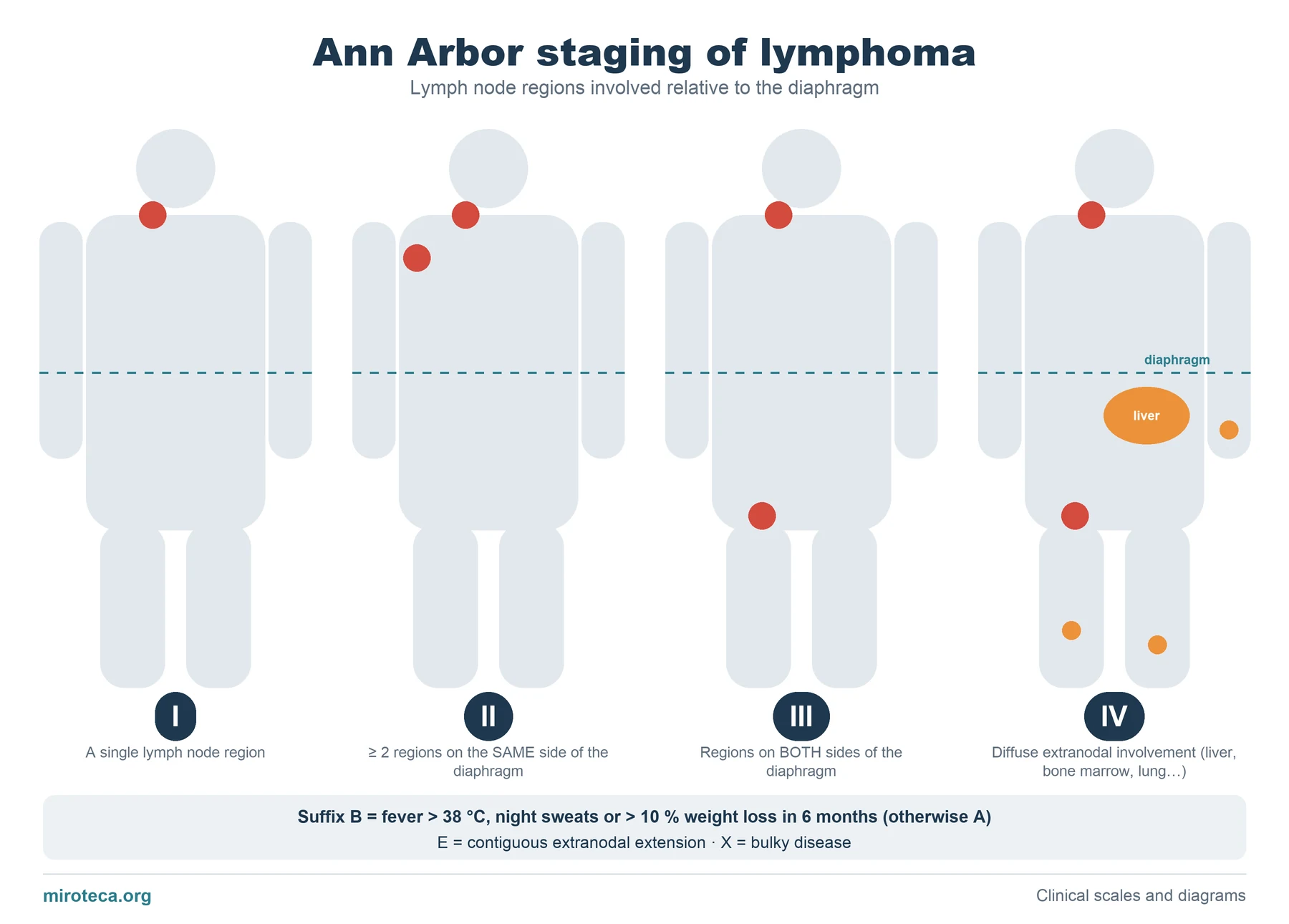 Ann Arbor staging of lymphoma: stages I to IV
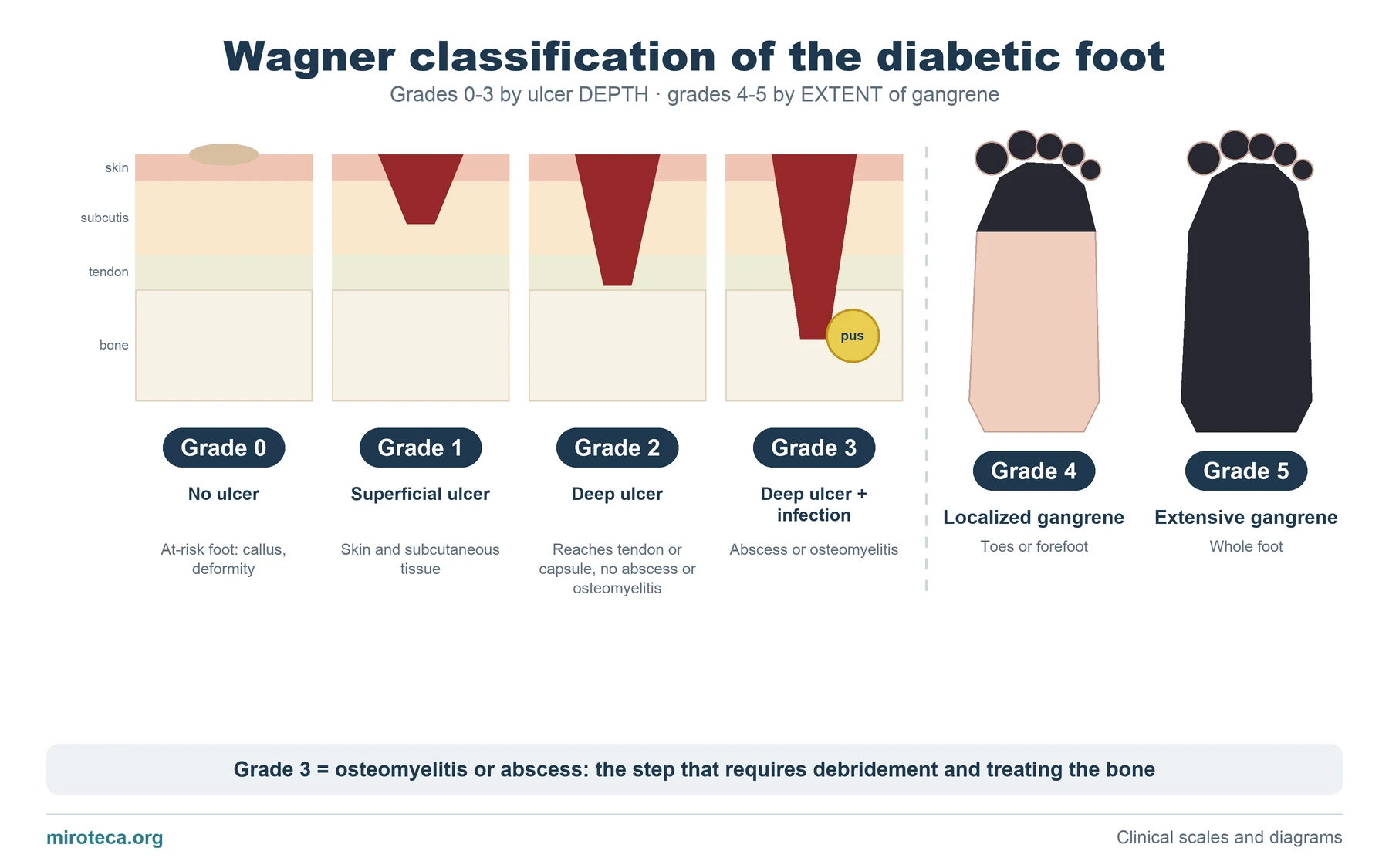 Wagner classification of diabetic foot ulcers: grades 0 to 5
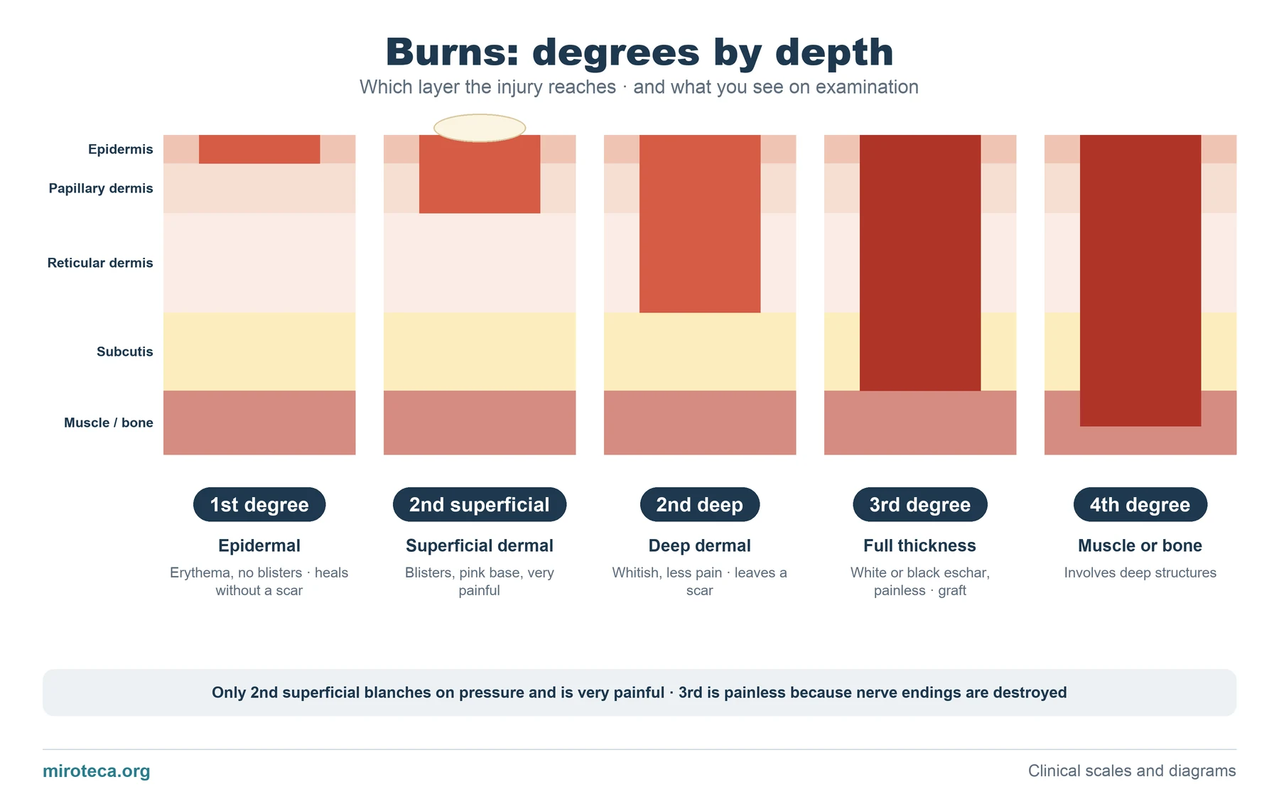 Burn degrees by depth: first to fourth degree
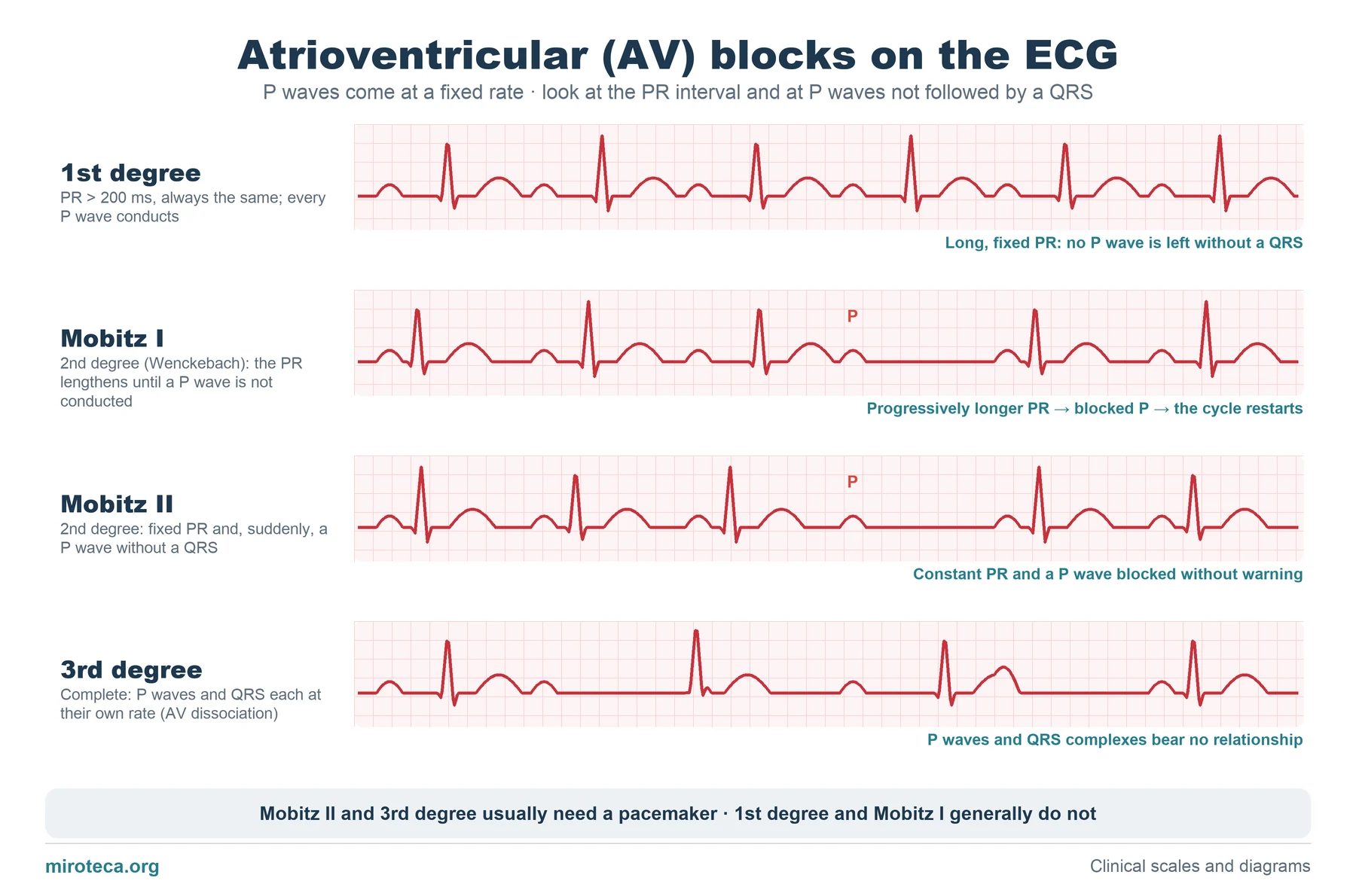 AV block on the ECG: first degree, Mobitz I, Mobitz II and complete
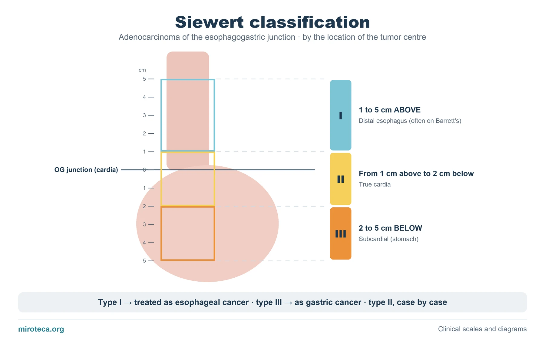 Siewert classification of esophagogastric junction adenocarcinoma
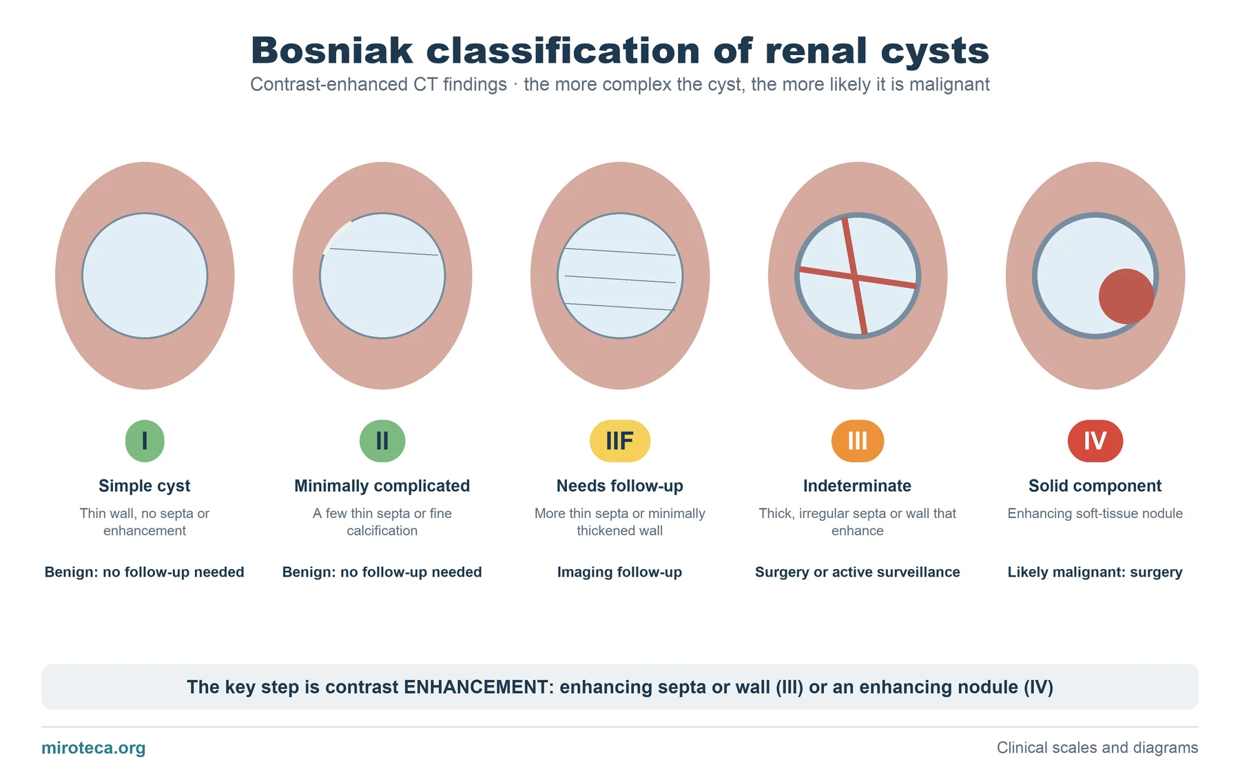 Bosniak classification of renal cysts: categories I to IV
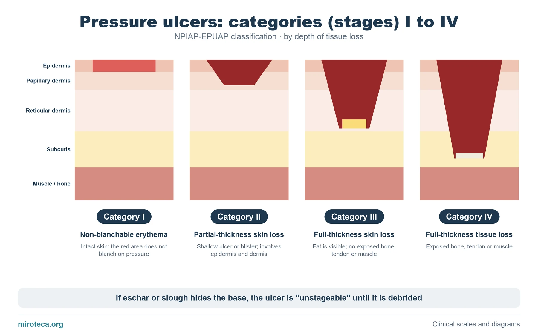 Pressure ulcer stages: categories I to IV
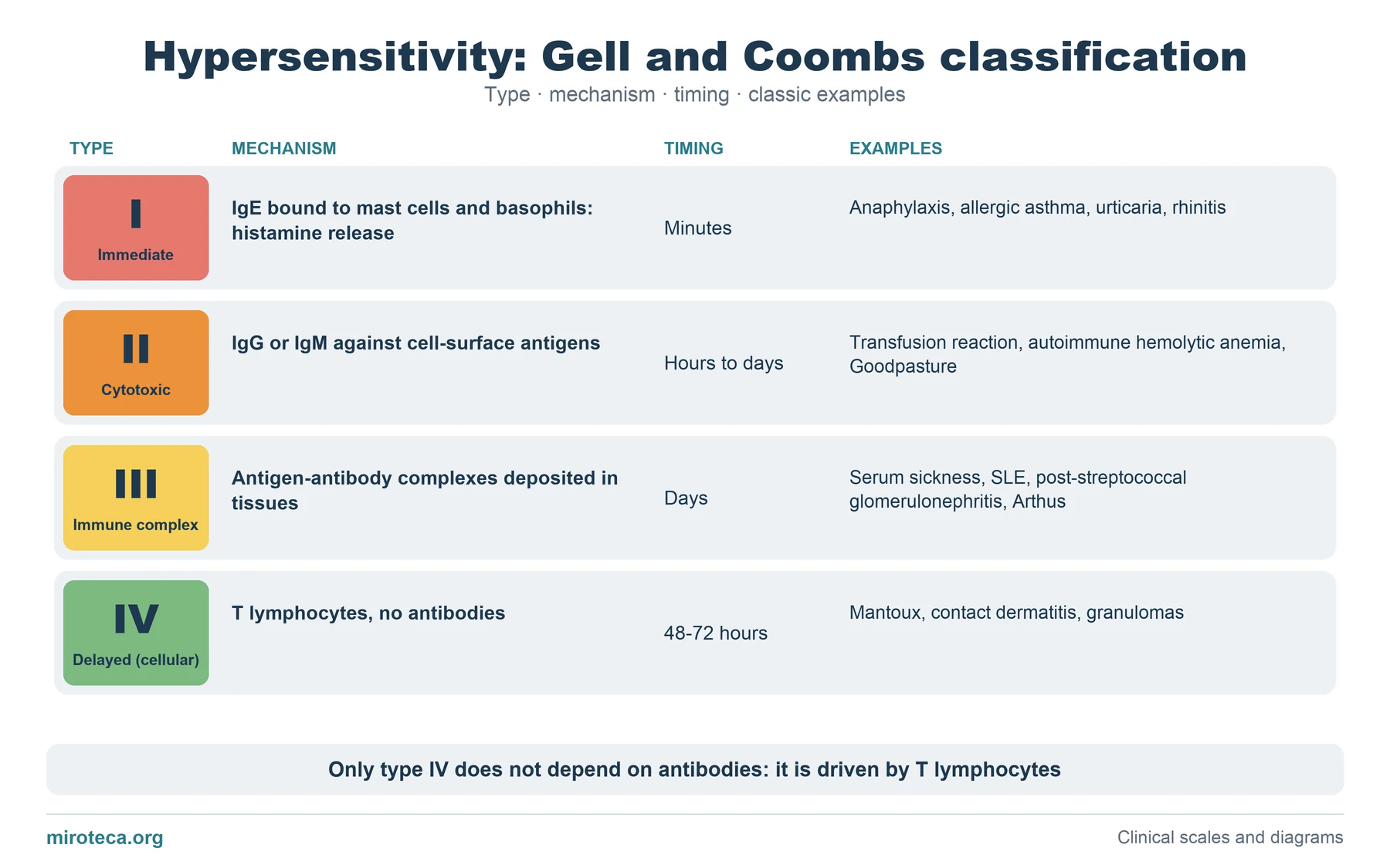 Gell and Coombs classification: the 4 types of hypersensitivity
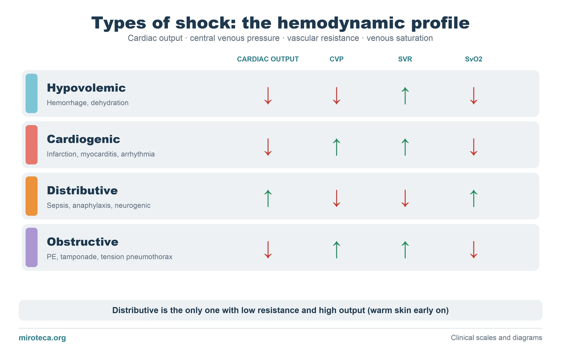 Types of shock: the hemodynamic profile
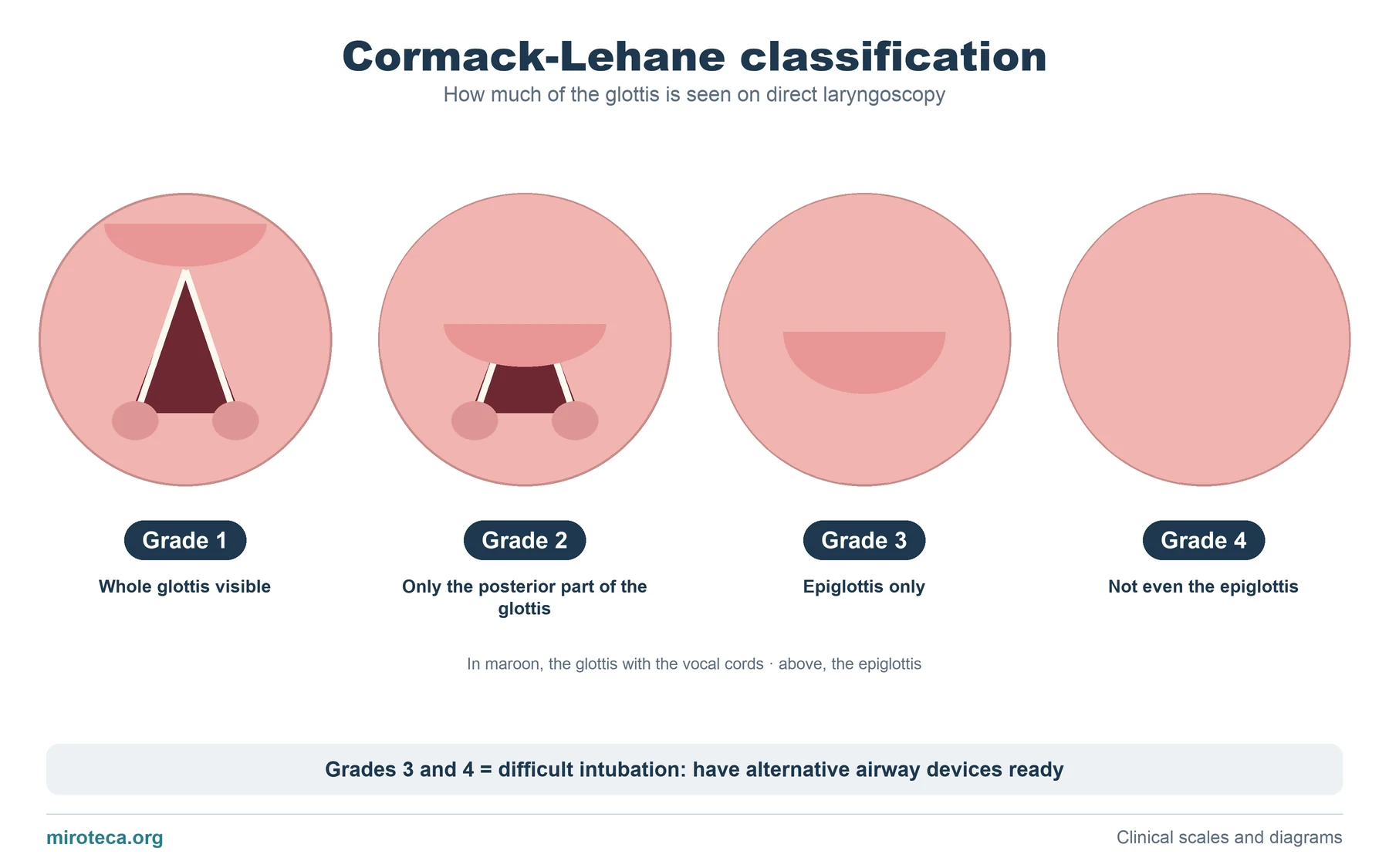 Cormack-Lehane classification of laryngoscopy: grades 1 to 4