
What the diagram shows
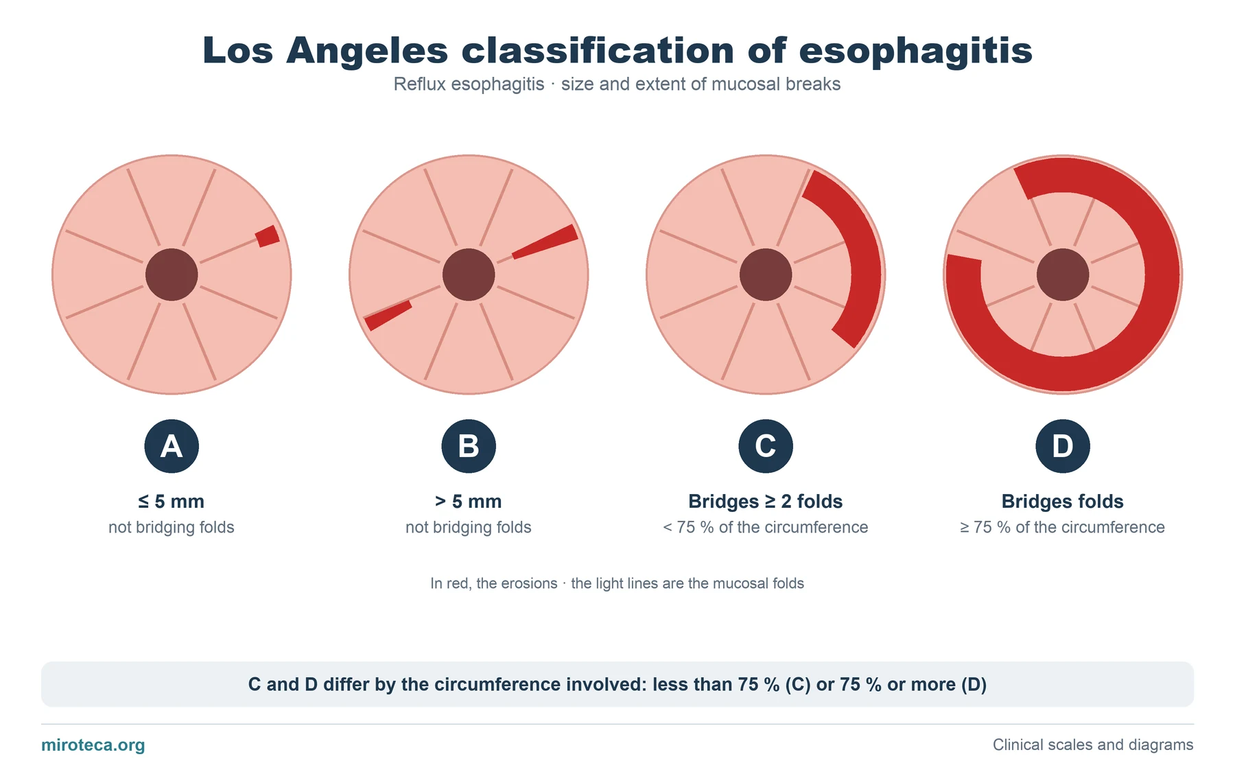
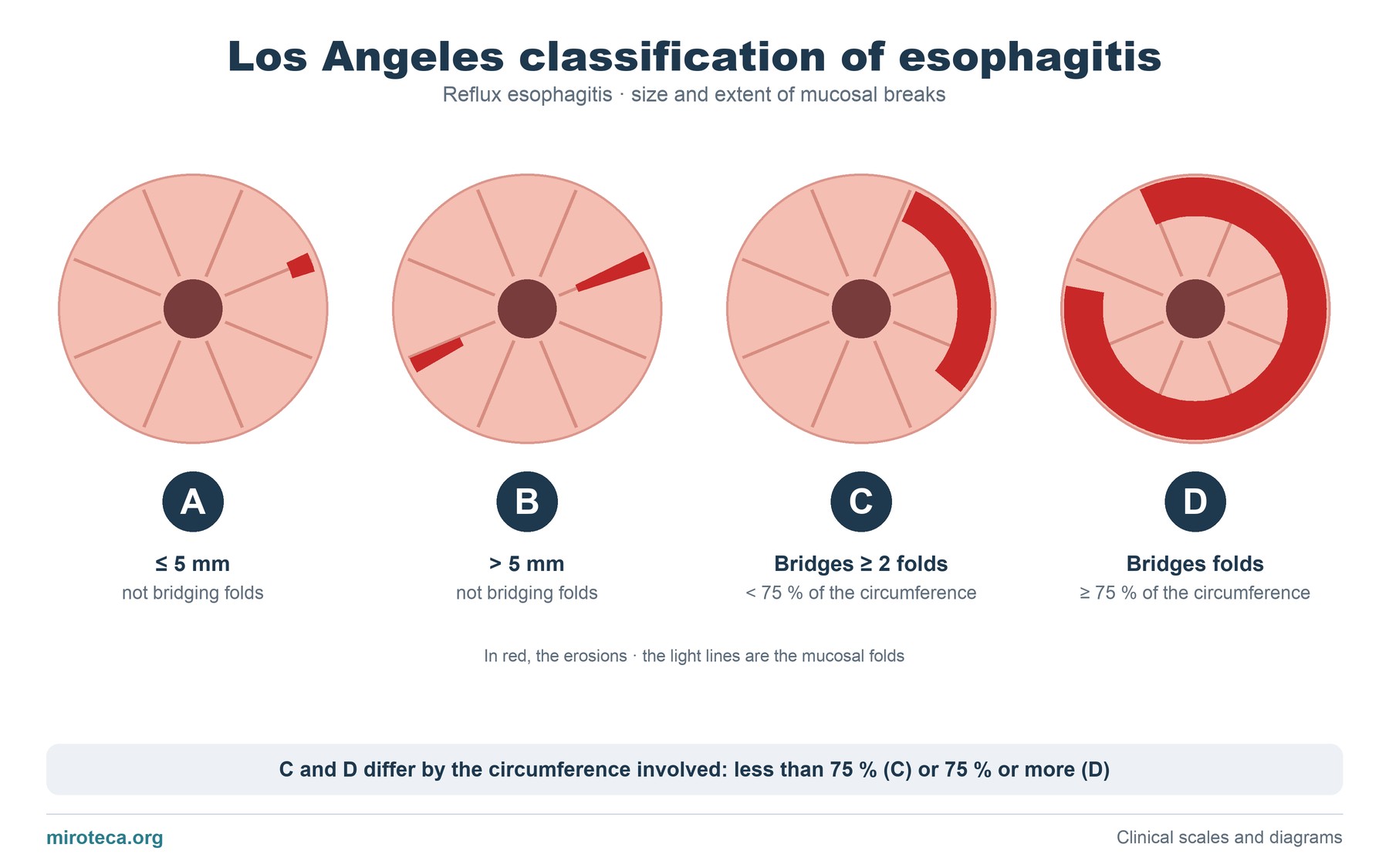
Each circle is the esophageal lumen seen through the endoscope, with the mucosal folds as spokes. The red areas are the mucosal breaks (erosions) caused by acid reflux.
From left to right the lesion grows: first in length (A and B) and then in extent around the esophagus (C and D). The classification does not measure depth, only the size and continuity of the breaks.
The detail that settles it
The key to going from B to C is whether the breaks are continuous between the tops of two or more folds. As long as each break stays on its own fold, however long, it is at most grade B. When they cross and join, it is C or D, and the difference between those two is set by 75 % of the circumference.
Grades C and D correspond to severe esophagitis and are associated with more complications (stricture, Barrett's esophagus).
Common mistakes
A common slip is confusing Los Angeles with the older Savary-Miller classification, which includes complications (ulcer, stricture, Barrett's) as grades of their own. Los Angeles only describes the mucosal breaks.
Another mistake is thinking that grade A on its own confirms reflux disease: small breaks can be seen in people without symptoms, and in mild grades confirmation usually needs other tests, such as pH monitoring.
In exam questions
Questions usually describe the endoscopic finding (for example, confluent erosions over less than three quarters of the circumference) and ask for the grade. Following the order of the diagram is enough: size, whether they are continuous, and how much circumference they cover.
Keep practicing
This page is the visual summary. To test yourself with real clinical reasoning, Medicinedle, the medical Wordle, gives you one clinical case a day: guess the diagnosis in six tries, with a new clue after every miss. It is free and available in English.