
What the diagram shows
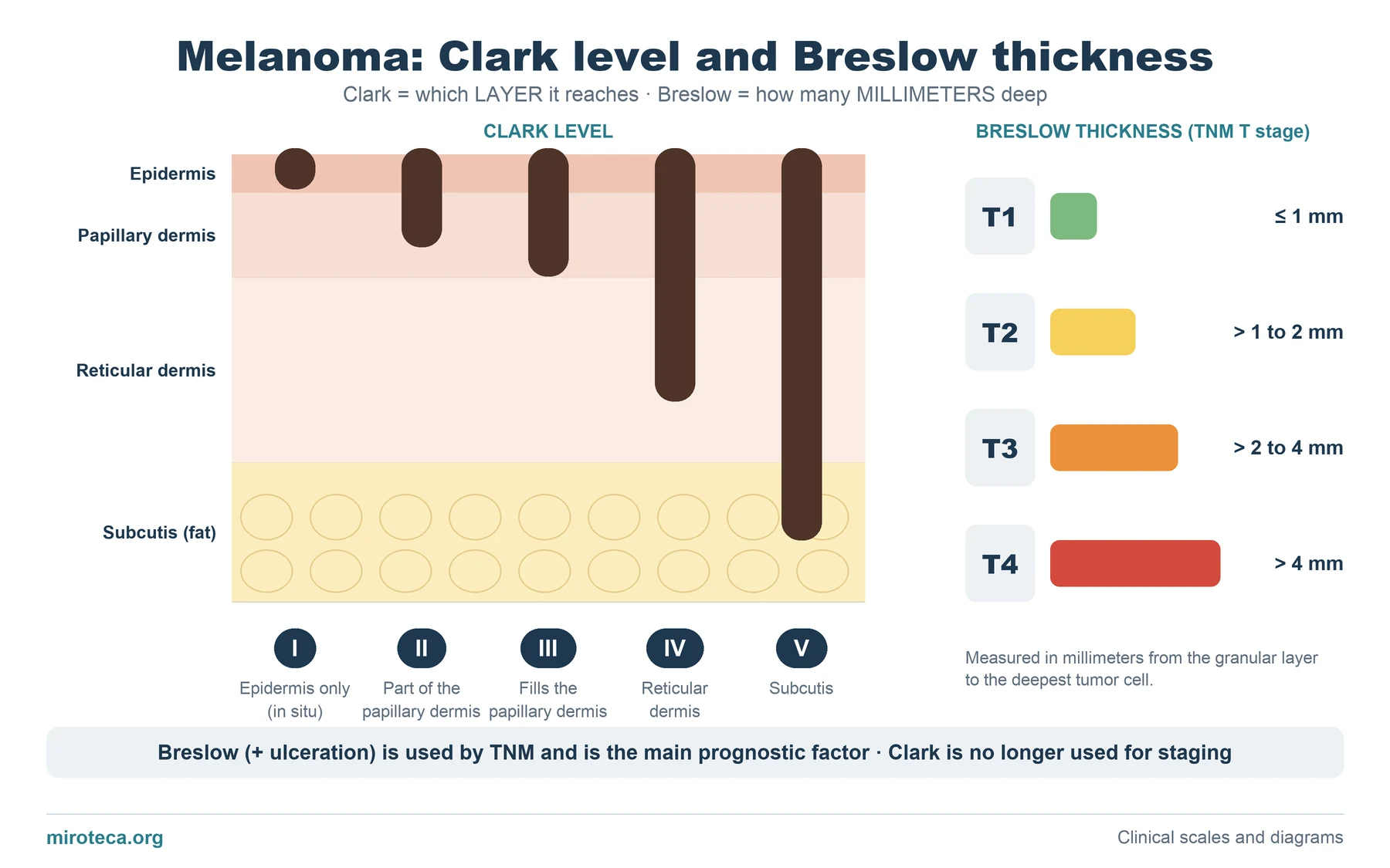
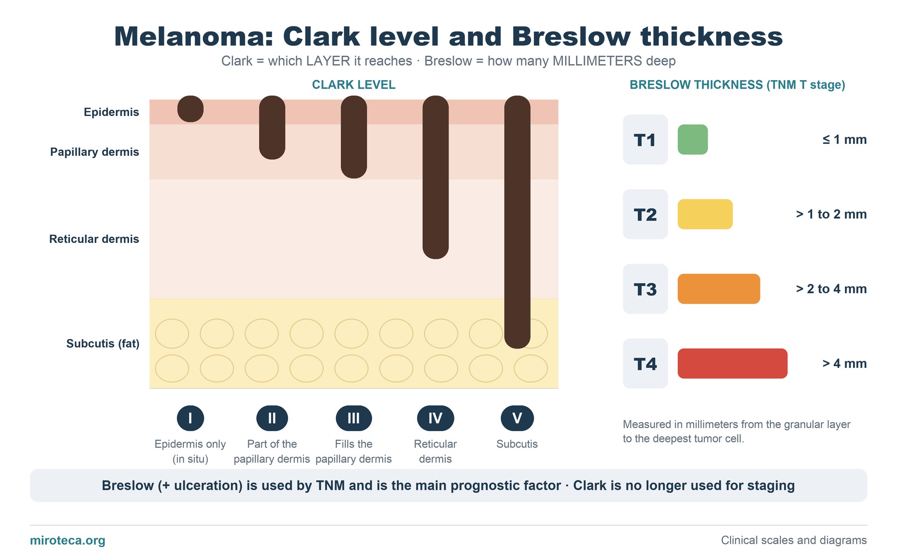
On the left, a skin section with its four layers: epidermis, papillary dermis, reticular dermis and subcutis. Each dark bar is a melanoma reaching a little deeper: these are the five Clark levels, which describe invasion by anatomical layer.
On the right, the Breslow thickness, which does not look at layers but at millimeters: it is measured from the granular layer of the epidermis to the deepest tumor cell. Its cut-offs (1, 2 and 4 mm) define the T category of the TNM system.
The detail that settles it
The two systems look as if they say the same thing, but they are not equivalent: skin thickness varies a lot between body sites. A 1 mm melanoma can reach the reticular dermis on the eyelid and stay in the papillary dermis on the back. That is why the millimeter is more reproducible than the layer, and why it prevailed.
Breslow thickness, together with ulceration, is the most important prognostic factor in localized melanoma. Clark level still appears in pathology reports by tradition, but current staging no longer uses it.
Common mistakes
The most frequent error is giving Clark level more prognostic weight than Breslow thickness, or mixing up the levels (Clark III is the whole papillary dermis, not the reticular dermis). Another is thinking that Breslow is measured from the skin surface: it is measured from the granular layer, and in an ulcerated lesion from the base of the ulcer.
Thickness is obtained from the excised specimen, which is why a suspicious lesion should be biopsied by excision whenever possible.
In exam questions
Questions usually give a pathology report with both figures and ask which one determines prognosis or management (margin re-excision, sentinel lymph node biopsy). The answer is almost always in the Breslow thickness and whether there is ulceration.
Keep practicing
This page is the visual summary. To test yourself with real clinical reasoning, Medicinedle, the medical Wordle, gives you one clinical case a day: guess the diagnosis in six tries, with a new clue after every miss. It is free and available in English.