
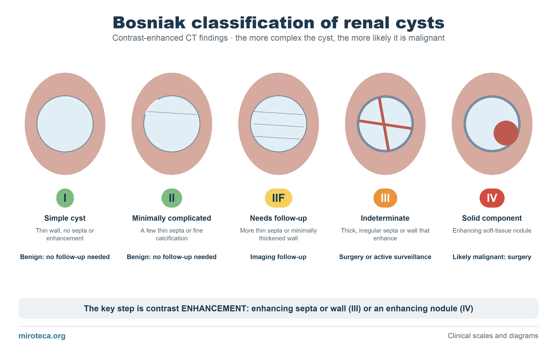
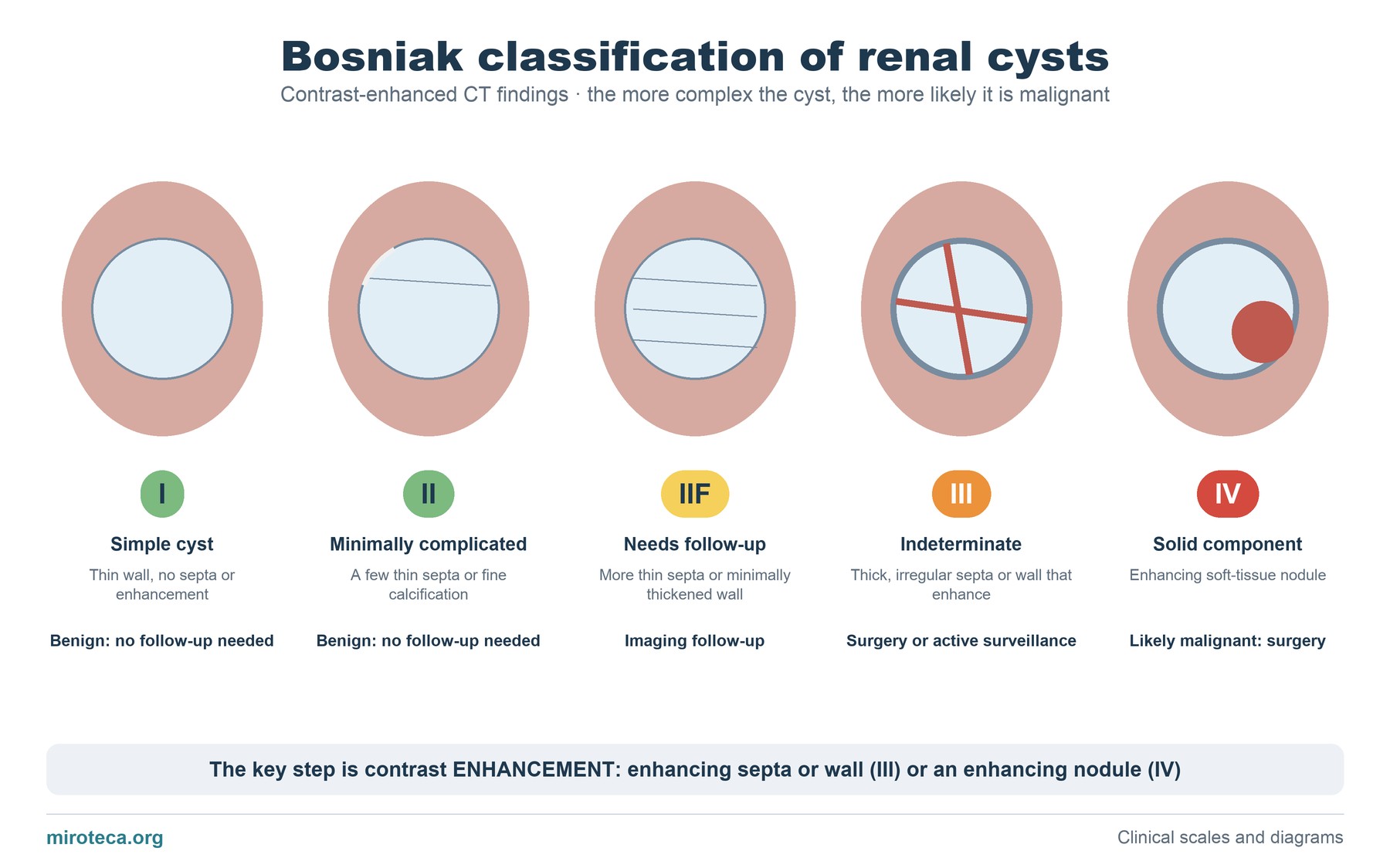
What the diagram shows
Each drawing is a kidney with a cyst that becomes more complex: first thin septa appear, then thick septa and, finally, a solid nodule. The Bosniak classification applies to renal cysts studied with contrast-enhanced CT (or MRI).
Below each one, in bold, the management: nothing for categories I and II, follow-up for IIF and consider surgery for III and IV.
The detail that settles it
The key word is enhancement: a wall, septum or nodule that takes up contrast has a blood supply, and that is what separates benign from suspicious. Categories III and IV are defined precisely by structures that enhance.
Category IIF (F for follow-up) covers cysts that are not simple but not clearly suspicious either; they are monitored with imaging to see whether they change.
Common mistakes
Classifying a cyst with ultrasound or with non-contrast CT: Bosniak needs contrast to assess enhancement. Another error is operating on every cyst with calcification: fine calcification is compatible with category II, which is benign.
An updated version published in 2019 refines the definitions (wall and septal thickness, nodules) and adds MRI criteria; the five categories are kept.
In exam questions
Questions usually describe the cyst on CT and ask for the category or the management. The key question is whether anything enhances and whether it is solid.
Keep practicing
This page is the visual summary. To test yourself with real clinical reasoning, Medicinedle, the medical Wordle, gives you one clinical case a day: guess the diagnosis in six tries, with a new clue after every miss. It is free and available in English.