
What the diagram shows
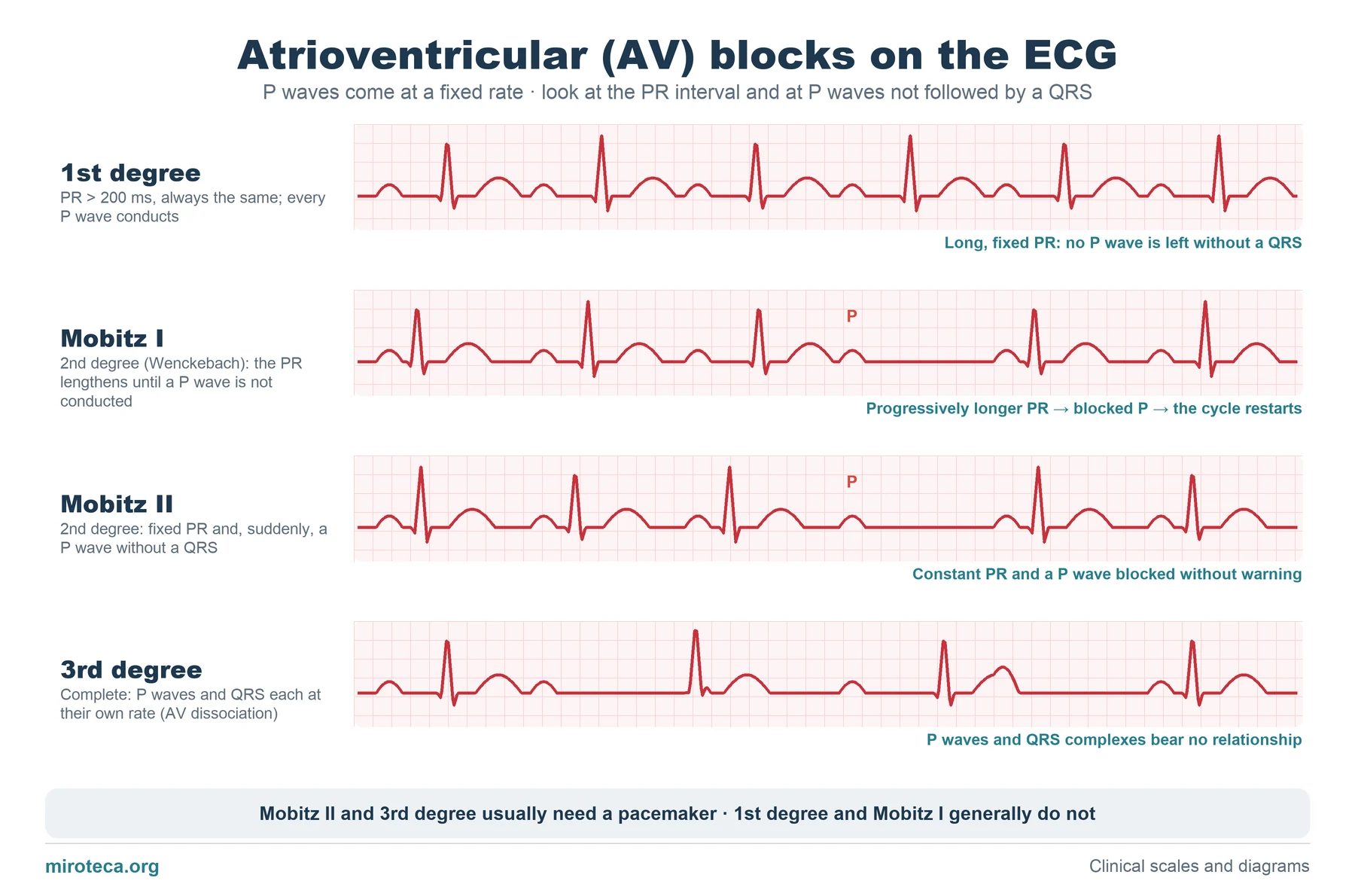
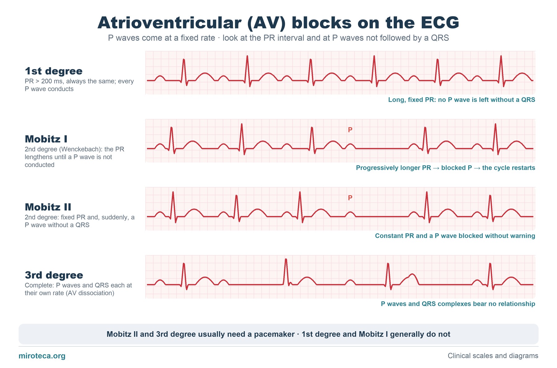
Four ECG strips in which the P waves appear at regular intervals, as in the real heart. What changes between them is the PR interval (from the P wave to the QRS) and whether a P wave is left without a QRS after it.
In the second-degree strips, the blocked P wave is marked with a letter "P".
The detail that settles it
To tell the two types of second-degree block apart, look at the PR of the beats that are conducted. In Mobitz I it lengthens progressively before the blocked P wave and shortens afterwards. In Mobitz II it is always the same and the block comes without warning.
That difference reflects where the problem is: Mobitz I is usually at the AV node and tends to be benign; Mobitz II is usually infra-Hisian, with a risk of progressing to complete block, which is why it usually warrants a pacemaker.
Common mistakes
With 2:1 conduction (one P wave conducts and the next does not) it is impossible to tell Mobitz I from II, because there are no two consecutive PR intervals to compare. Another error is calling any bradycardia with P waves "complete block": in complete block P waves and QRS complexes are fully independent and the PR changes with every beat without any pattern.
Reversible causes should not be forgotten before indicating a pacemaker: drugs that slow the node (beta-blockers, calcium channel blockers, digoxin), hyperkalemia or an inferior myocardial infarction.
In exam questions
Questions usually give an ECG or its description and ask for the type of block and the management. First check whether every P wave is conducted; then whether the PR changes.
Keep practicing
This page is the visual summary. To test yourself with real clinical reasoning, Medicinedle, the medical Wordle, gives you one clinical case a day: guess the diagnosis in six tries, with a new clue after every miss. It is free and available in English.