
What the diagram shows
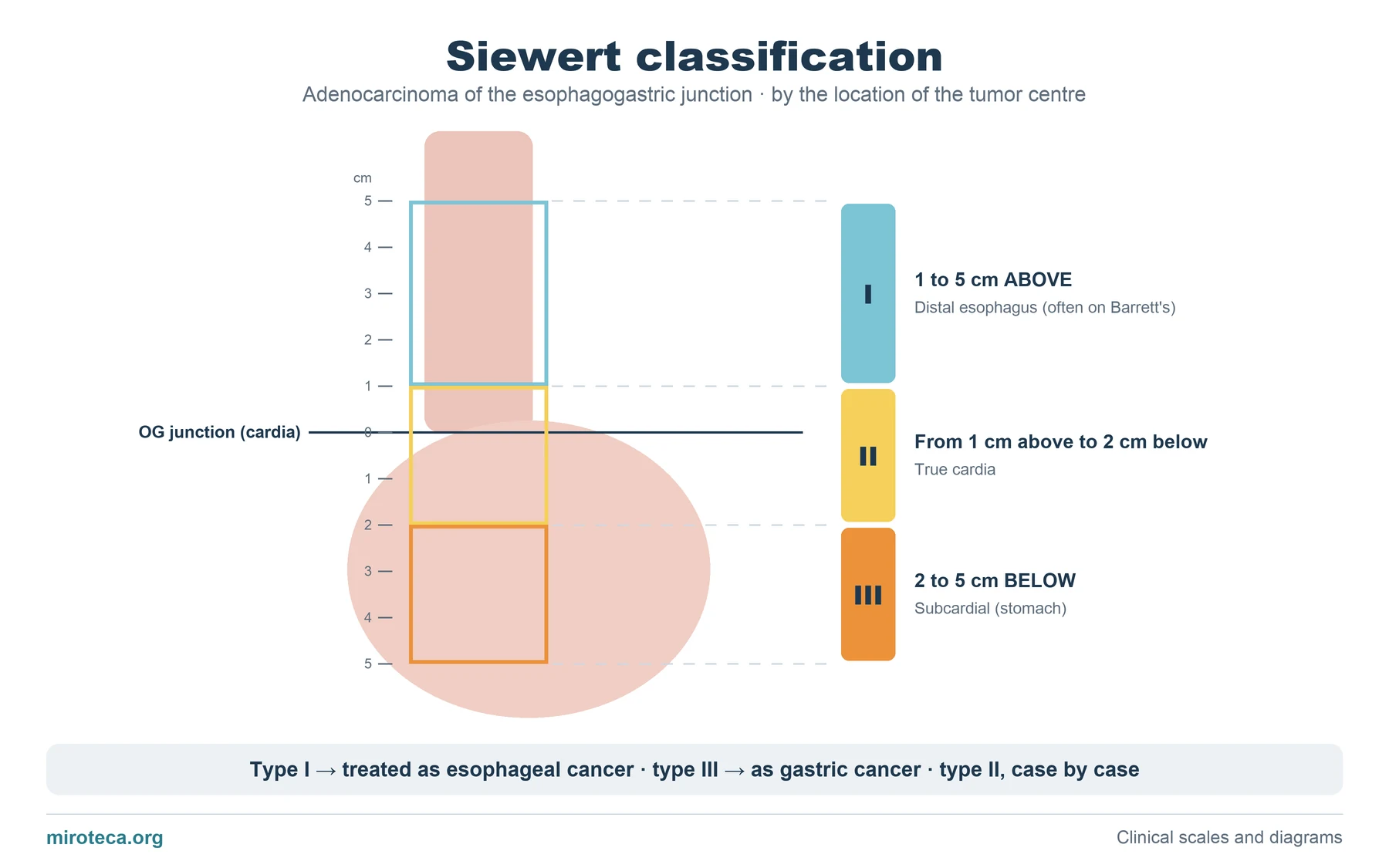
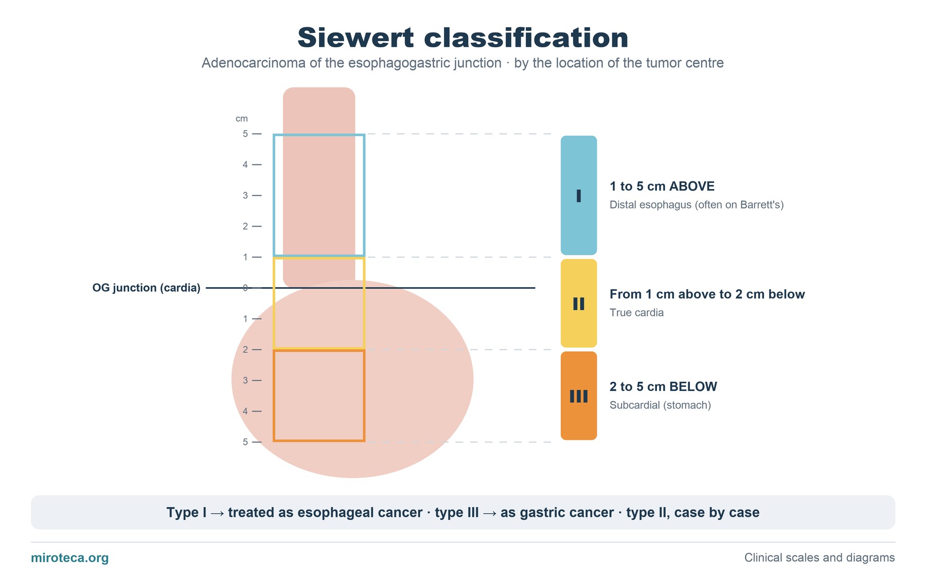
The distal esophagus opens into the stomach at the esophagogastric junction (the horizontal line). The ruler on the left marks centimeters above and below that line, and the three colored rectangles are the zones of each type.
Siewert classifies adenocarcinoma of that region by where the center of the tumor lies, not its edge.
The detail that settles it
The classification is used to plan surgery. Type I behaves like distal esophageal cancer, often arising on Barrett's esophagus, and is treated as such (esophagectomy). Type III behaves like gastric cancer and is treated with gastrectomy. Type II sits in between, and the technique depends on its extent.
Current TNM staging follows a similar criterion: tumors whose center lies 2 cm or less from the cardia and that involve it are staged as esophageal.
Common mistakes
The most common slip is applying Siewert to squamous cell carcinoma: the classification is only for adenocarcinoma of the junction. Another is measuring from the edge of the tumor instead of its center.
The esophagogastric junction should not be confused with the diaphragmatic hiatus: in a hiatal hernia they can be several centimeters apart.
In exam questions
Questions usually give the distance from the tumor to the cardia at endoscopy and ask for the type or the indicated surgery. The three bands of the diagram solve it.
Keep practicing
This page is the visual summary. To test yourself with real clinical reasoning, Medicinedle, the medical Wordle, gives you one clinical case a day: guess the diagnosis in six tries, with a new clue after every miss. It is free and available in English.