
What the diagram shows
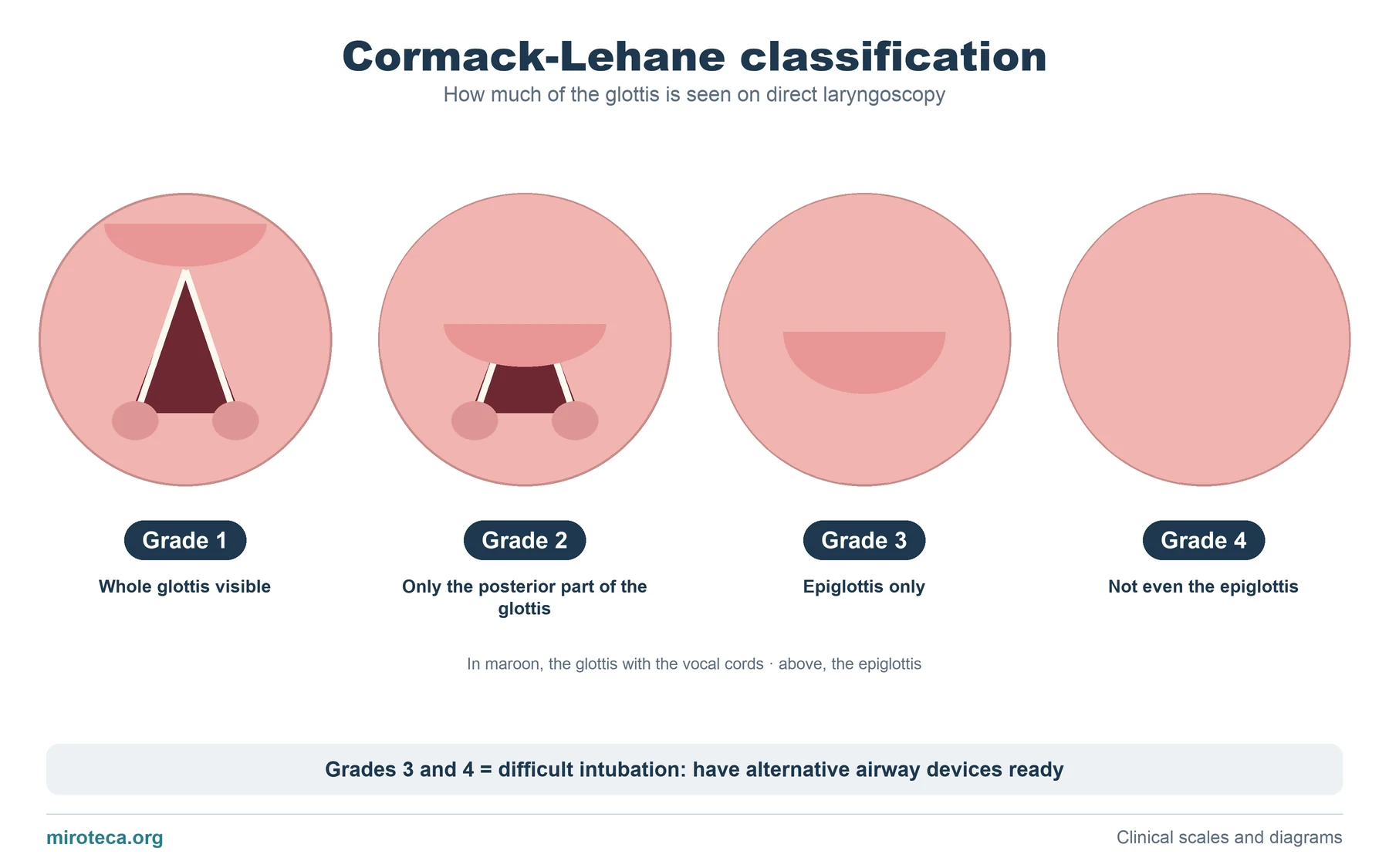
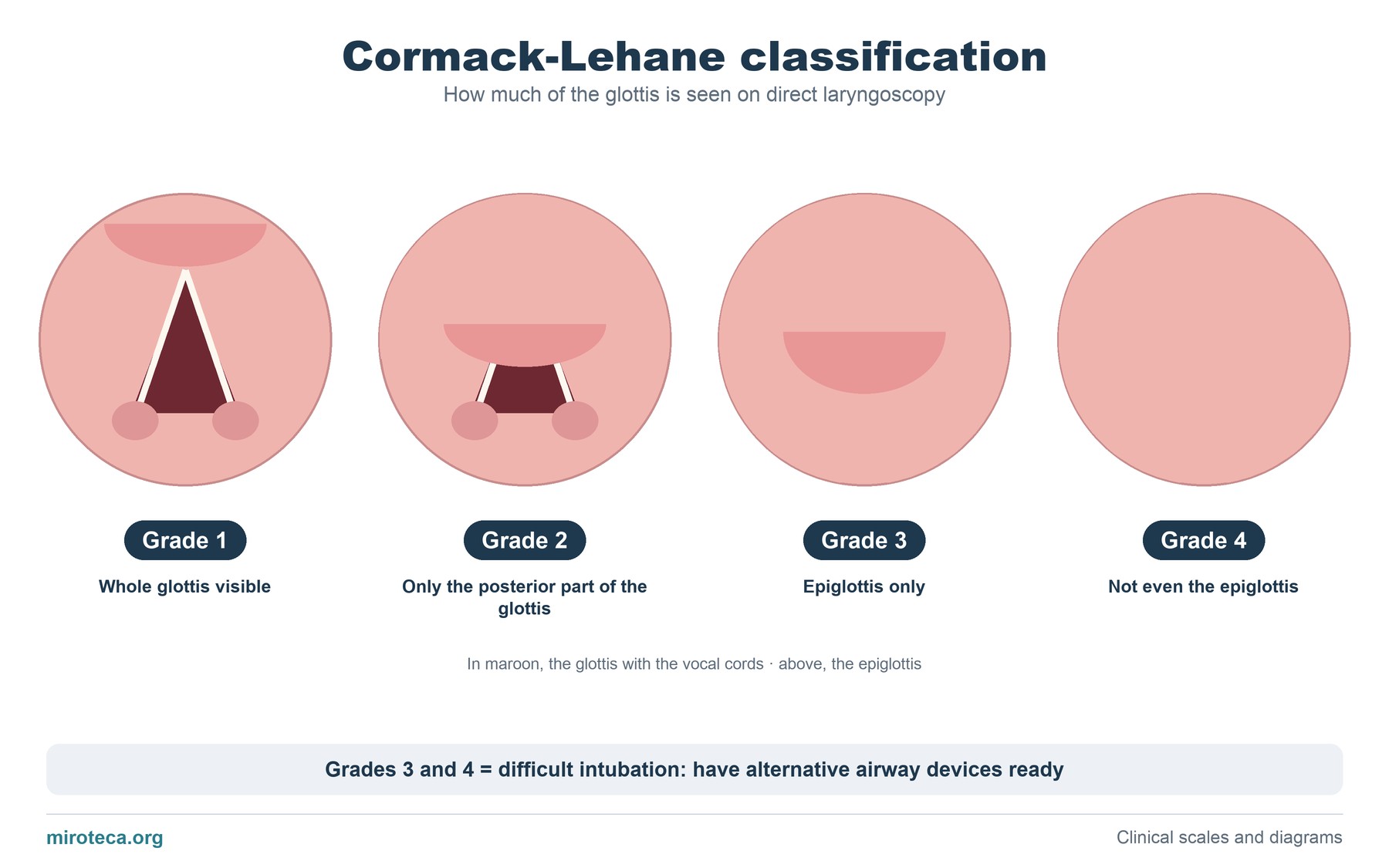
Each circle is what the anesthetist sees through the laryngoscope. At the top is the epiglottis; below it, the glottis (in maroon) with the vocal cords in an inverted "V", and at the bottom, the arytenoids.
From left to right the view is hidden from the front: first the anterior part of the glottis disappears, then the whole glottis, and finally the epiglottis as well.
The detail that settles it
Cormack-Lehane is obtained during laryngoscopy, whereas Mallampati is assessed beforehand, with the patient sitting and the mouth open. Mallampati predicts; Cormack-Lehane confirms what is actually seen.
Grades 3 and 4 define a difficult laryngoscopy: they require alternative devices to be ready (stylet or bougie, videolaryngoscope, laryngeal mask) and a plan if intubation fails.
Common mistakes
A frequent error is mixing up the two middle grades: in grade 2 part of the glottis (its posterior part) is still visible, whereas in grade 3 only the epiglottis is seen. Another is assuming that a low Mallampati guarantees an easy laryngoscopy: the correlation between the two is imperfect.
There are also modified versions that split grade 2 into 2a and 2b or grade 3 into 3a and 3b; exams usually use the original four-grade classification.
In exam questions
Questions usually describe what is seen at intubation and ask for the grade, or ask which scale is done before (Mallampati) and which during (Cormack-Lehane).
Keep practicing
This page is the visual summary. To test yourself with real clinical reasoning, Medicinedle, the medical Wordle, gives you one clinical case a day: guess the diagnosis in six tries, with a new clue after every miss. It is free and available in English.