
What the diagram shows
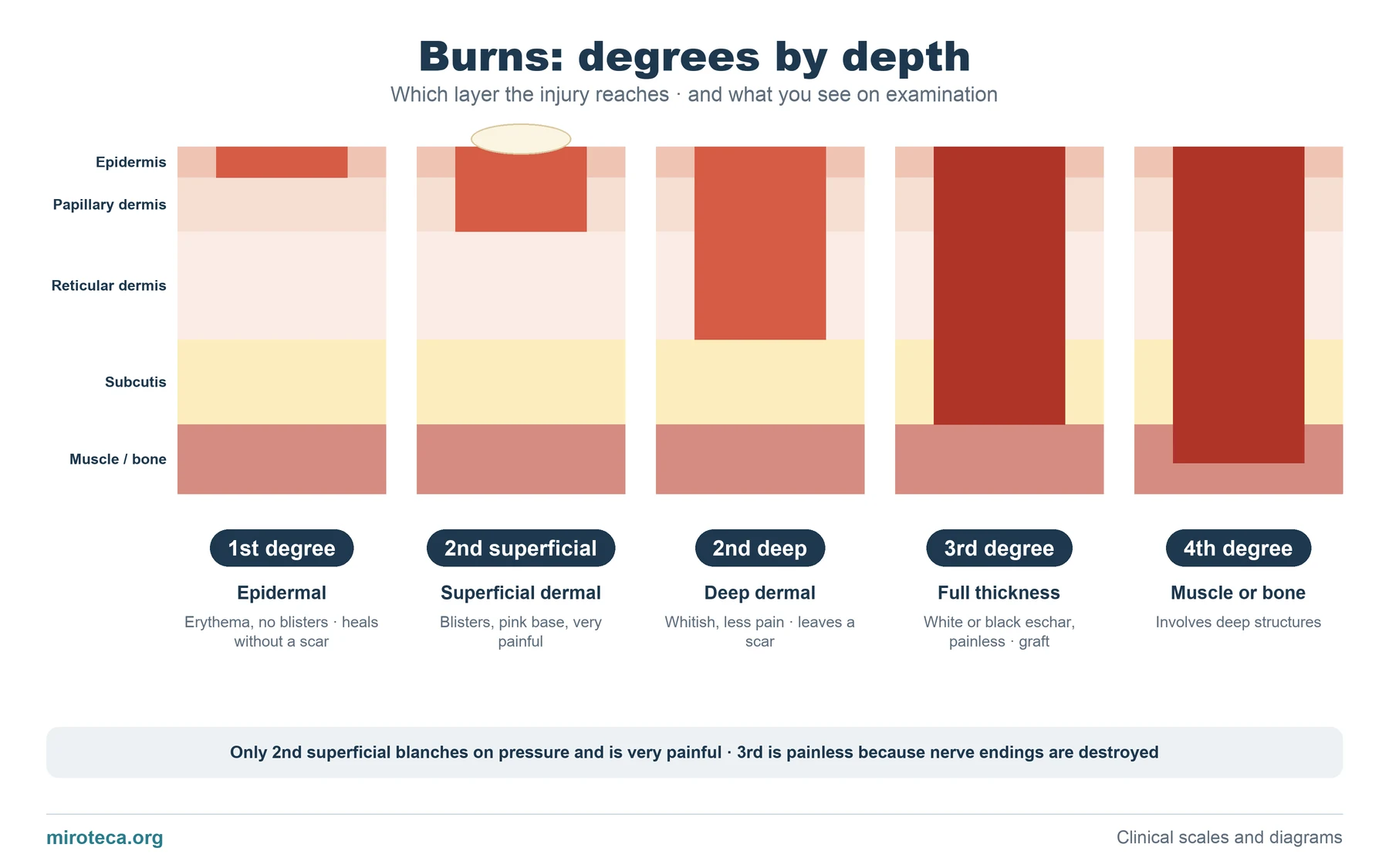
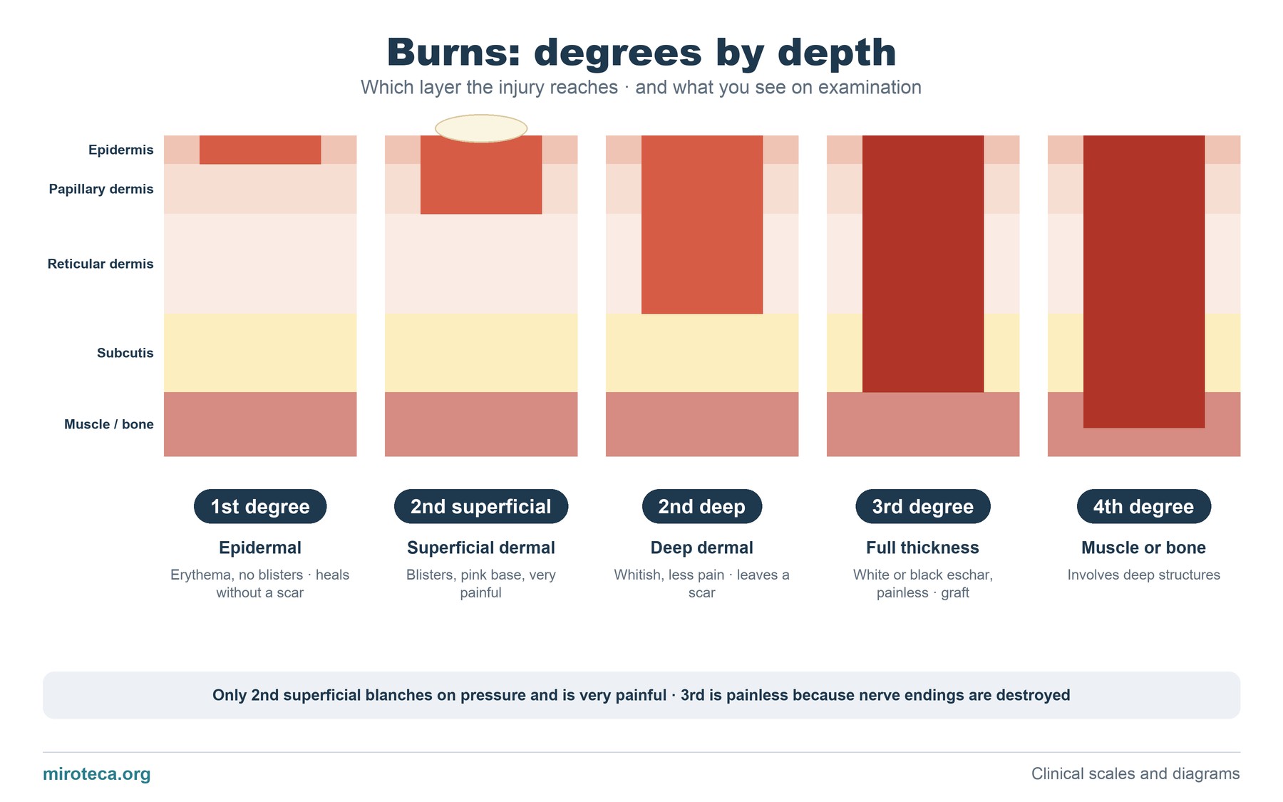
Five skin sections with their layers (epidermis, papillary dermis, reticular dermis, subcutis and muscle or bone). The red area is the damaged tissue, which goes deeper from left to right.
Current terminology speaks of epidermal, superficial dermal, deep dermal and full-thickness burns; the classic "degrees" are still the most used in practice and in exams.
The detail that settles it
What separates a burn that heals on its own from one that needs surgery is whether skin appendages (follicles and glands) remain in the dermis, from which the skin regenerates. In superficial dermal burns many remain and healing takes about two weeks; in deep dermal burns few remain, healing takes more than three weeks and leaves a scar, so grafting is often needed.
Examination helps: superficial dermal burns are very painful, have blisters and the base blanches on pressure; full-thickness burns are painless, because nerve endings are destroyed, and do not blanch.
Common mistakes
A frequent error is being reassured because a burn does not hurt: absence of pain suggests depth, not mildness. Another is estimating depth only on the first day; burns can deepen over the first 48-72 hours, which is why they are reassessed.
First-degree burns are not included when calculating the total body surface area burned or the fluid replacement.
In exam questions
Questions describe the appearance (blisters, color, pain, blanching) and ask for the depth or the management: conservative care for superficial burns, consider grafting for deep dermal and full-thickness burns.
Keep practicing
This page is the visual summary. To test yourself with real clinical reasoning, Medicinedle, the medical Wordle, gives you one clinical case a day: guess the diagnosis in six tries, with a new clue after every miss. It is free and available in English.