
What the chart shows
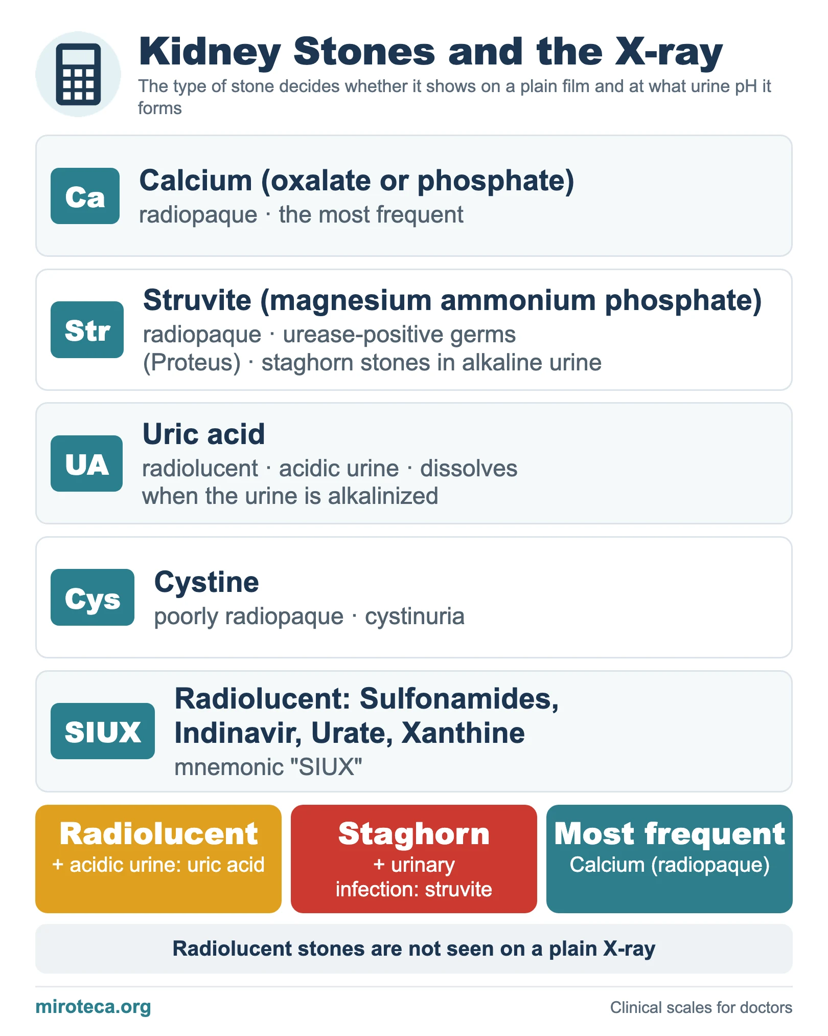
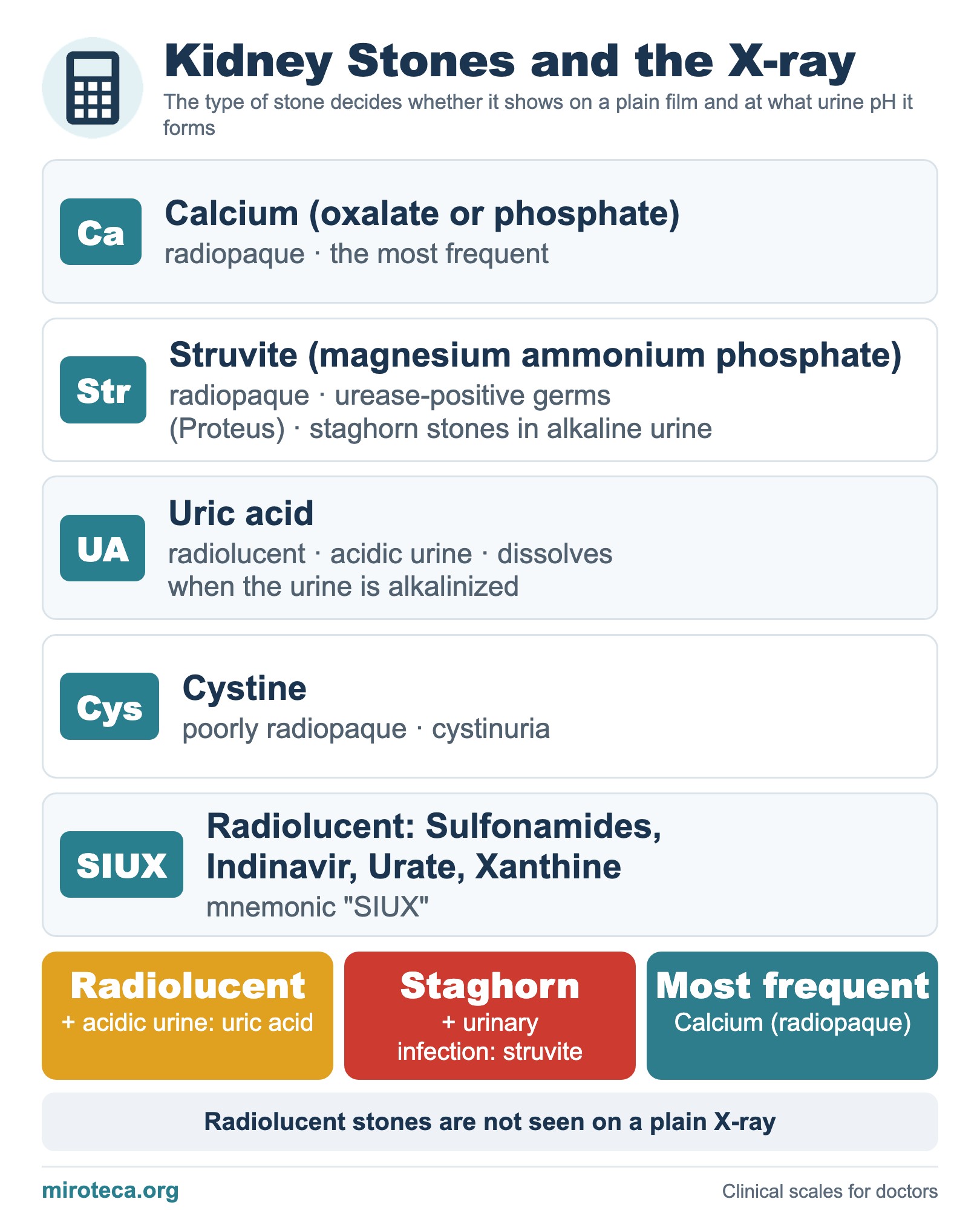
The type of kidney stone decides whether it can be seen on a plain X-ray and at what pH it forms, two data that help to guide diagnosis and prevention. The chart lists each type with its appearance, urine pH and typical setting.
The detail that settles it
Calcium oxalate and phosphate stones are the most frequent and radiopaque. Struvite stones are associated with infection by urease-positive germs (Proteus), are radiopaque and form staghorn stones in alkaline urine. Uric acid stones are radiolucent and form in acidic urine. Cystine stones are poorly radiopaque and appear in cystinuria.
Radiolucent plus acidic urine means uric acid (it dissolves by alkalinizing the urine). A staghorn stone with a urinary infection means struvite.
In exam questions
Short rules: the most frequent is calcium (radiopaque); a staghorn stone with infection is struvite; radiolucent stones are remembered with the mnemonic SIUX. See also uric acid.
Keep practicing
This page is the visual summary. To test yourself with real clinical reasoning, Medicinedle, a Wordle-inspired game, gives you one clinical case a day: guess the diagnosis in six tries, with a new clue after every miss. It is free and available in English.