
What the chart shows
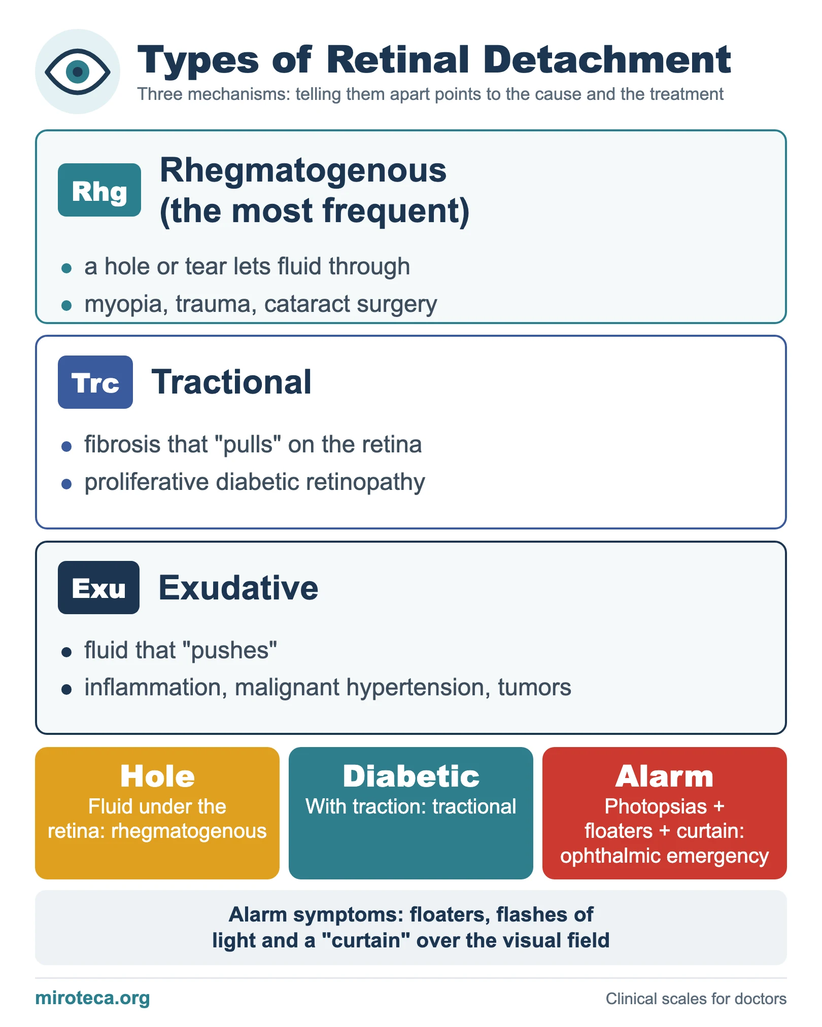
Retinal detachment has three mechanisms, and telling them apart points to the cause and the treatment. The chart gives what happens in each and its typical causes.
The detail that settles it
Rhegmatogenous detachment is the most frequent: a break or hole lets fluid pass under the retina (factors: myopia, trauma, cataract surgery). Tractional detachment is due to fibrosis that "pulls" on the retina, typical of proliferative diabetic retinopathy. Exudative detachment occurs when fluid "pushes" in inflammation, malignant hypertension or tumors.
The alarm picture is floaters, flashes of light (photopsias) and a "curtain" in the visual field: an ophthalmic emergency.
In exam questions
Short rules: a hole plus fluid means rhegmatogenous (the most common); a diabetic patient with traction means tractional; flashes, floaters and a curtain are an eye emergency.
Keep practicing
This page is the visual summary. To test yourself with real clinical reasoning, Medicinedle, a Wordle-inspired game, gives you one clinical case a day: guess the diagnosis in six tries, with a new clue after every miss. It is free and available in English.