
What the chart shows
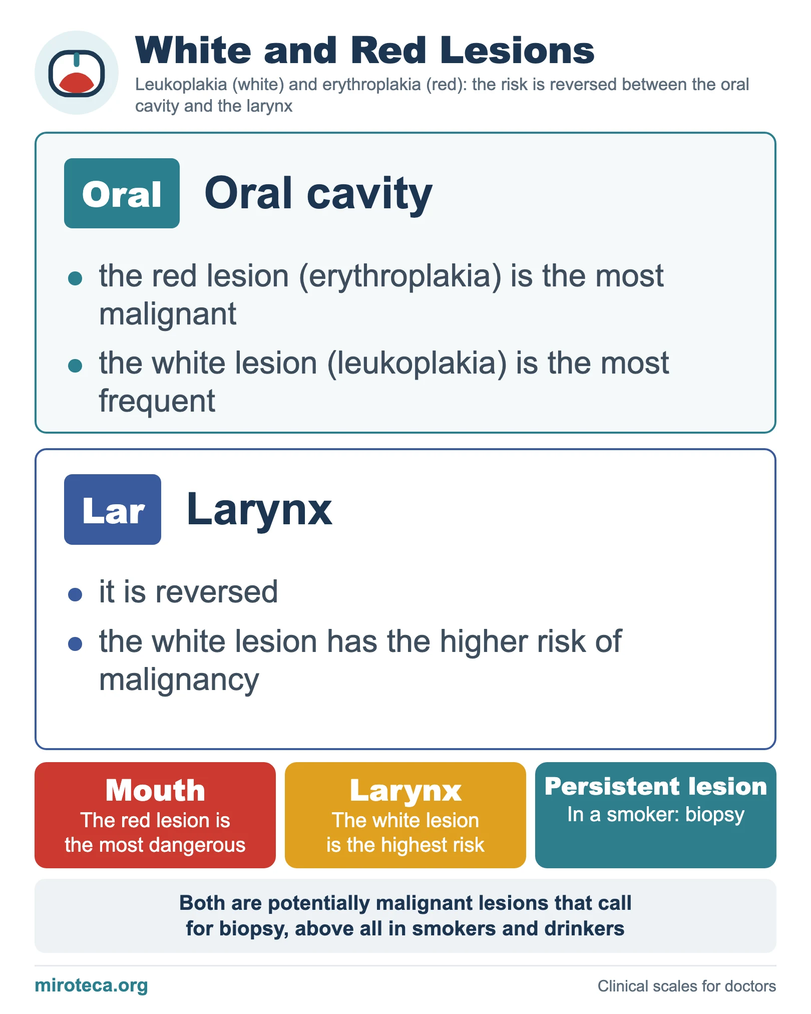
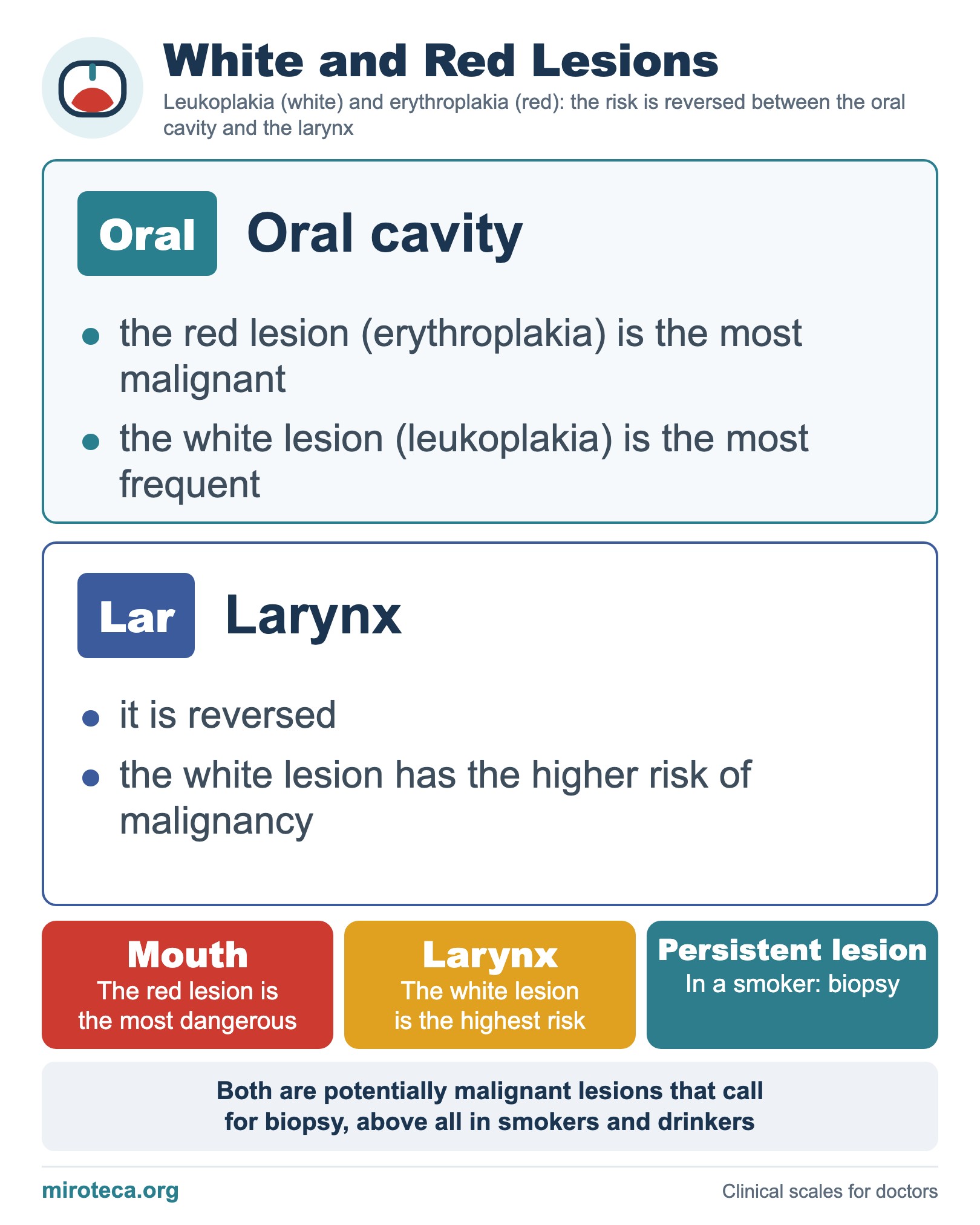
White and red lesions of the mucosa can be premalignant, and their risk is reversed between the oral cavity and the larynx. The chart sets the two sites side by side.
The detail that settles it
In the oral cavity, erythroplakia (red) is the most malignant, although leukoplakia (white) is the most frequent. In the larynx it is reversed: the white lesion has the higher risk of becoming malignant. Both are potentially malignant lesions that call for biopsy, above all in smokers and drinkers.
In exam questions
Short rules: in the mouth, the red lesion is the most dangerous; in the larynx, the white lesion is the highest risk; any persistent lesion in the mucosa of a smoker should be biopsied.
Keep practicing
This page is the visual summary. To test yourself with real clinical reasoning, Medicinedle, a Wordle-inspired game, gives you one clinical case a day: guess the diagnosis in six tries, with a new clue after every miss. It is free and available in English.