
What the chart shows
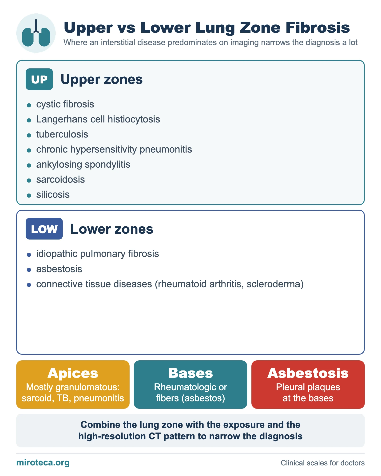
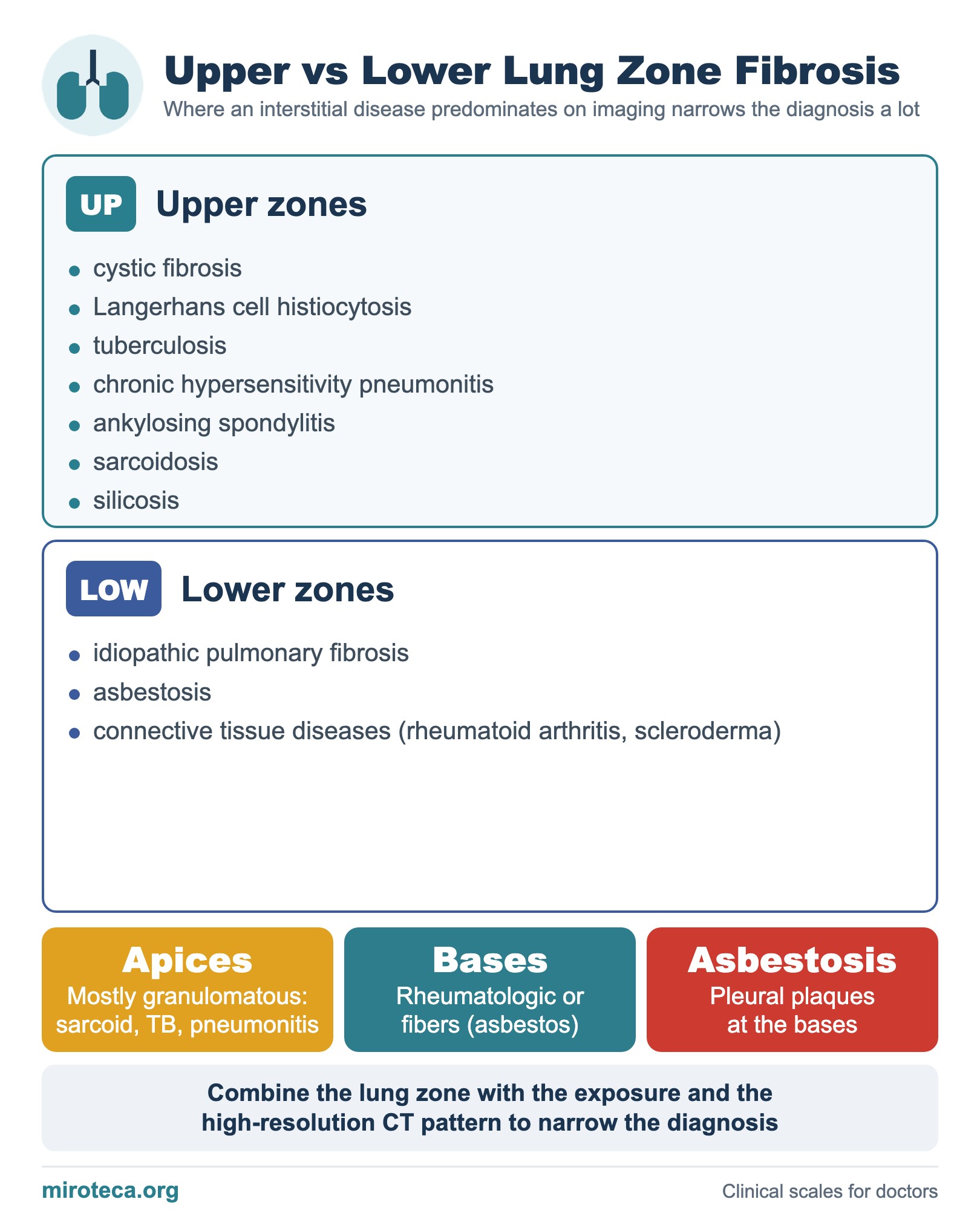
Whether an interstitial lung disease predominates in the upper or lower zones on imaging narrows the diagnosis a lot. The chart groups the ones that "go up" and the ones that prefer the bases.
The rule and its counterpart
Upper zones: cystic fibrosis, Langerhans cell histiocytosis, tuberculosis, chronic hypersensitivity pneumonitis, ankylosing spondylitis, sarcoidosis and silicosis. Lower zones: idiopathic pulmonary fibrosis, asbestosis and connective tissue diseases (rheumatoid arthritis, scleroderma).
An exam trick: almost all the "basal" diseases have a rheumatologic component or come from fibers (asbestos), while the "apical" ones are usually granulomatous (sarcoidosis, tuberculosis, pneumonitis). Tuberculosis and sarcoidosis can leave cavities or retractile fibrosis at the apices, while asbestosis comes with pleural plaques at the bases. See interstitial lung disease clues.
In exam questions
Short rules: idiopathic pulmonary fibrosis and asbestosis mean lower zones; sarcoidosis, silicosis and tuberculosis mean upper zones; combine the zone with the exposure and the CT pattern.
Keep practicing
This page is the visual summary. To test yourself with real clinical reasoning, Medicinedle, a Wordle-inspired game, gives you one clinical case a day: guess the diagnosis in six tries, with a new clue after every miss. It is free.
Prefer quick questions? The clinical scores quiz gives you 5 case-based questions in about a minute, with the answer explained.