What the chart shows
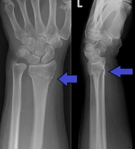
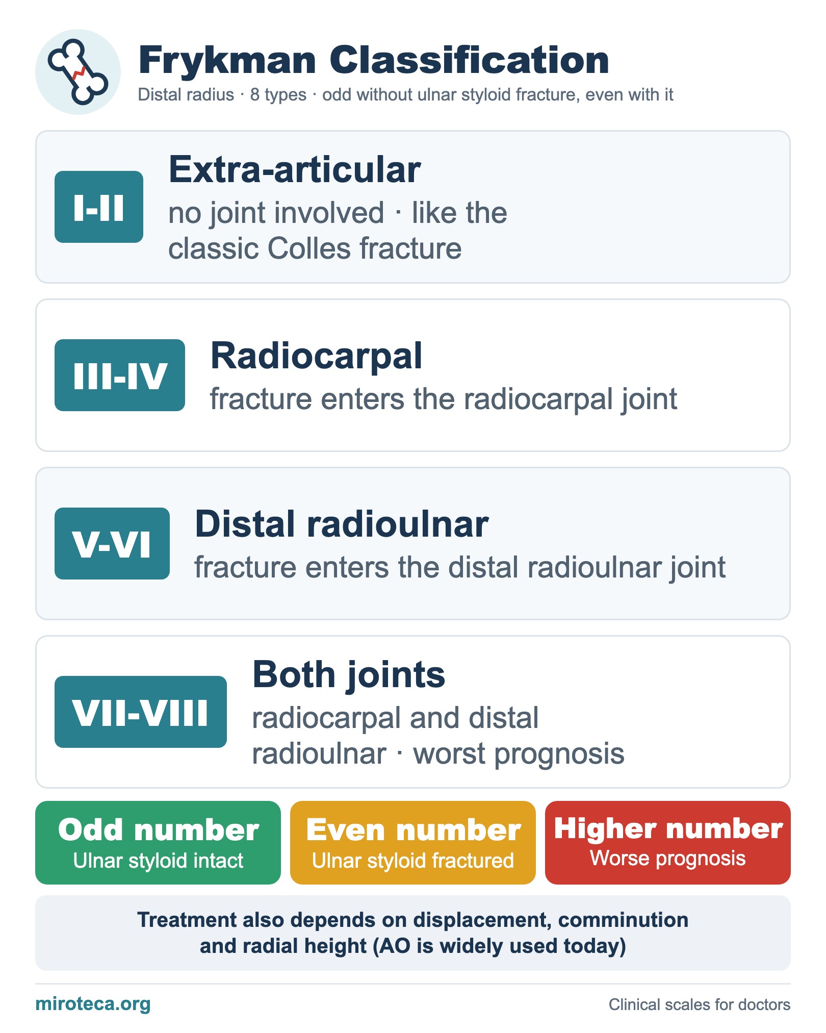
The Frykman classification sorts distal radius fractures (Colles, Smith and similar) into eight types by whether the fracture enters the radiocarpal joint, the distal radioulnar joint or both, and whether the ulnar styloid is also fractured.
The detail that settles it
Odd numbers have an intact ulnar styloid and even numbers a fractured one. Types I-II are extra-articular, III-IV involve the radiocarpal joint, V-VI the distal radioulnar joint and VII-VIII both. The higher the number, the worse the prognosis.
In exam questions
Frykman has prognostic value but does not define stability: treatment depends on displacement, dorsal comminution, radial shortening and age. Many surgeons now use the AO system (A extra-articular, B partial articular, C complete articular).
Keep practicing
This page is the visual summary. To test yourself with real clinical reasoning, Medicinedle, a Wordle-inspired game, gives you one clinical case a day: guess the diagnosis in six tries, with a new clue after every miss. It is free.
Prefer quick questions? The clinical scores quiz gives you 5 case-based questions in about a minute, with the answer explained.