What the chart shows
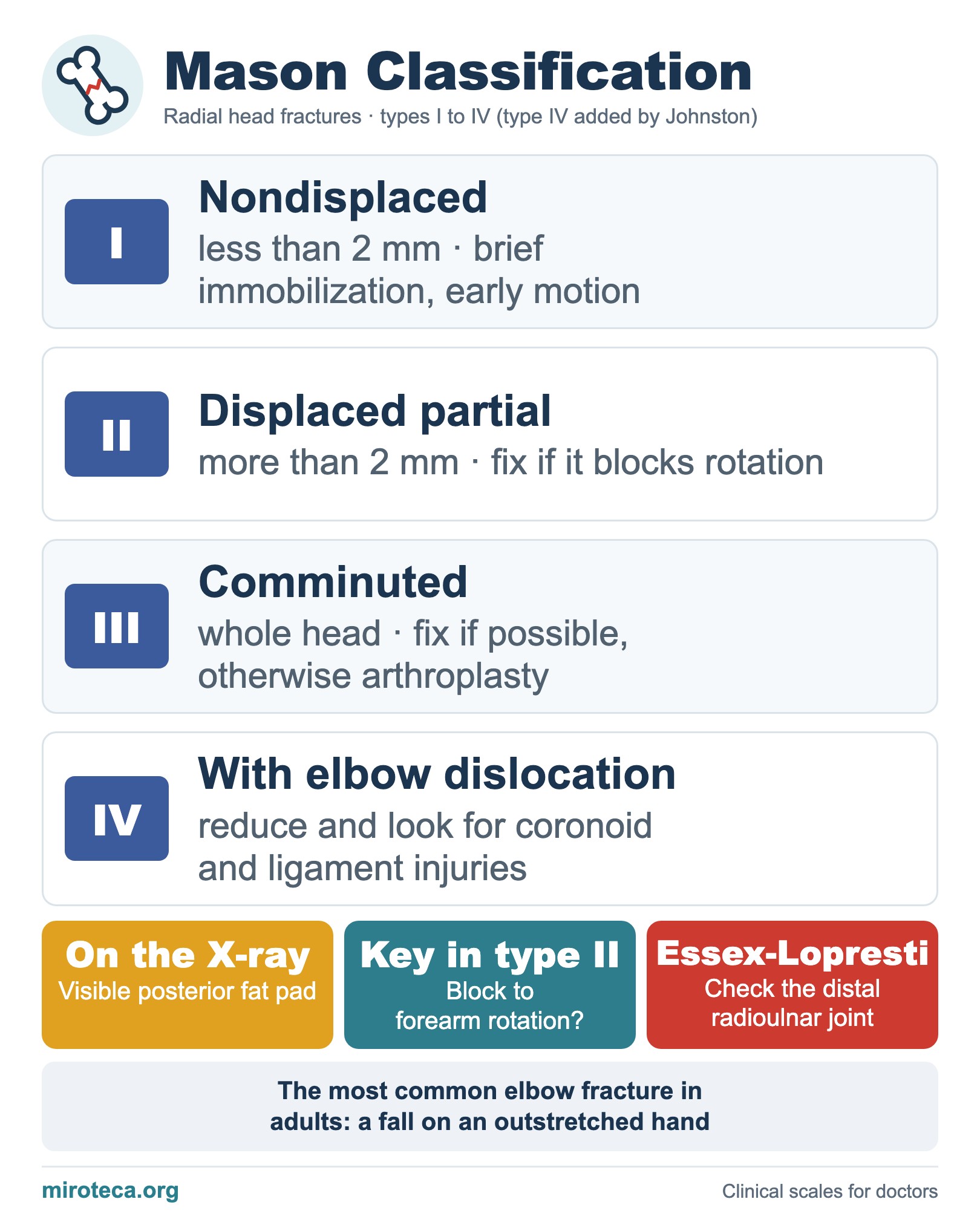
The Mason classification describes radial head fractures, the most common elbow fractures in adults, by displacement and comminution. Mason described three types in 1954; Johnston added type IV in 1962 for fractures with an elbow dislocation.
The detail that settles it
Type I (displaced less than 2 mm) does well with early motion; prolonged immobilization causes stiffness. In type II the deciding factor is a mechanical block to pronation and supination. Type III is comminuted and, if it cannot be fixed, radial head arthroplasty is considered.
In exam questions
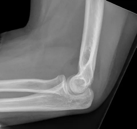
A visible posterior fat pad on an elbow X-ray means an effusion and an occult fracture until proven otherwise. The Essex-Lopresti injury combines a radial head fracture, interosseous membrane tear and distal radioulnar joint dislocation, so forearm stability must be checked before excising the radial head.
Keep practicing
This page is the visual summary. To test yourself with real clinical reasoning, Medicinedle, a Wordle-inspired game, gives you one clinical case a day: guess the diagnosis in six tries, with a new clue after every miss. It is free.
Prefer quick questions? The clinical scores quiz gives you 5 case-based questions in about a minute, with the answer explained.