What the chart shows
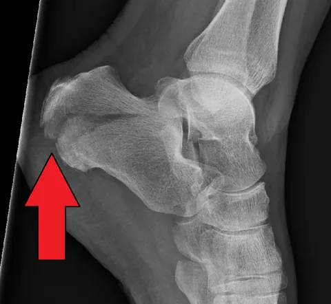
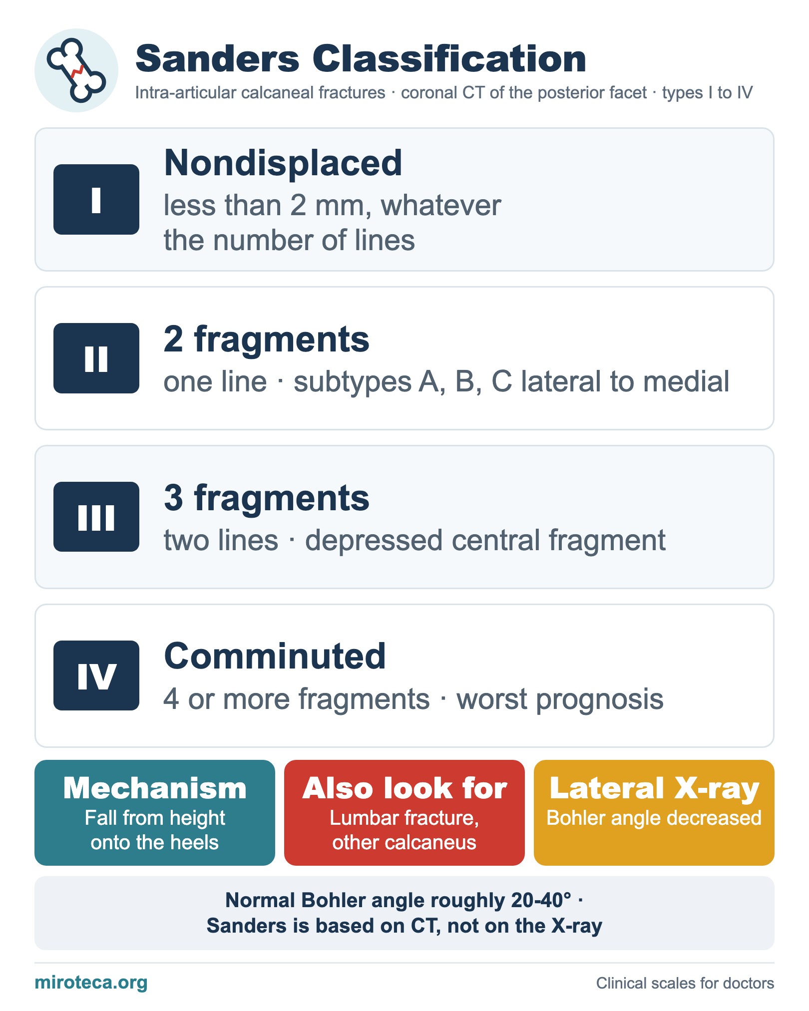
The Sanders classification describes intra-articular calcaneal fractures on CT: on the coronal image where the posterior facet is widest, the articular fragments are counted. The calcaneus is the most commonly fractured tarsal bone, usually after a fall from height onto the heels.
The detail that settles it
Type I is nondisplaced (less than 2 mm) whatever the number of lines; type II has two fragments, type III three (often with a depressed central fragment) and type IV four or more. Types II and III are subdivided into A, B and C by the position of the line, lateral to medial. More fragments mean a worse prognosis.
In exam questions
On the lateral X-ray, Bohler angle (normal roughly 20-40 degrees) decreases when the posterior facet is depressed. With a fall from height, look for a lumbar spine fracture and a fracture of the other calcaneus.
Keep practicing
This page is the visual summary. To test yourself with real clinical reasoning, Medicinedle, a Wordle-inspired game, gives you one clinical case a day: guess the diagnosis in six tries, with a new clue after every miss. It is free.
Prefer quick questions? The clinical scores quiz gives you 5 case-based questions in about a minute, with the answer explained.