What the chart shows
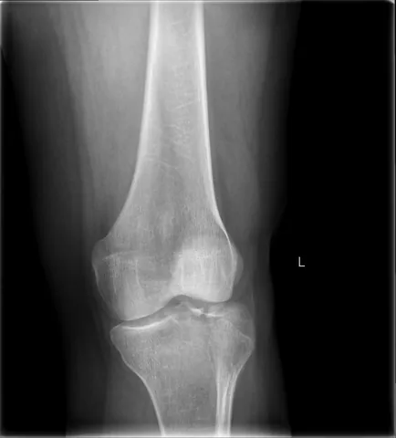
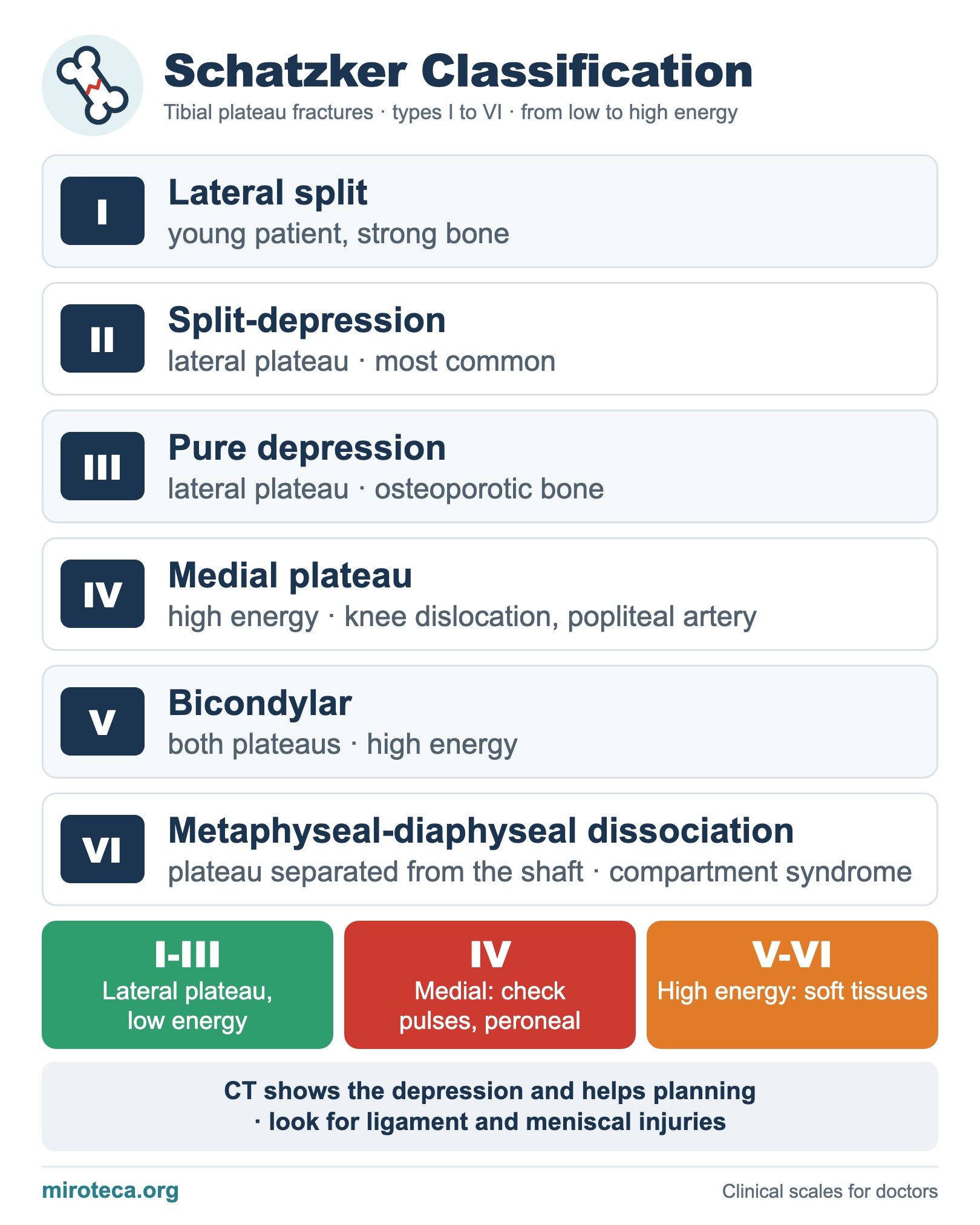
The Schatzker classification divides tibial plateau fractures into six types. Types I-III involve the lateral plateau and are usually low energy; types IV-VI are high energy and more often come with soft tissue, ligament and vascular injuries. CT is almost always used to assess depression and plan surgery.
The detail that settles it
A useful rule: type I is the young patient (strong bone that splits), type III the older patient (soft bone that depresses) and type II, the most common, does both. Type IV, the medial plateau fracture, can be a knee dislocation equivalent.
In exam questions
In types IV-VI, check distal pulses and the ankle-brachial index, watch the peroneal nerve and monitor for compartment syndrome. Severe fractures are often temporized with an external fixator until the soft tissues settle.
Keep practicing
This page is the visual summary. To test yourself with real clinical reasoning, Medicinedle, a Wordle-inspired game, gives you one clinical case a day: guess the diagnosis in six tries, with a new clue after every miss. It is free.
Prefer quick questions? The clinical scores quiz gives you 5 case-based questions in about a minute, with the answer explained.