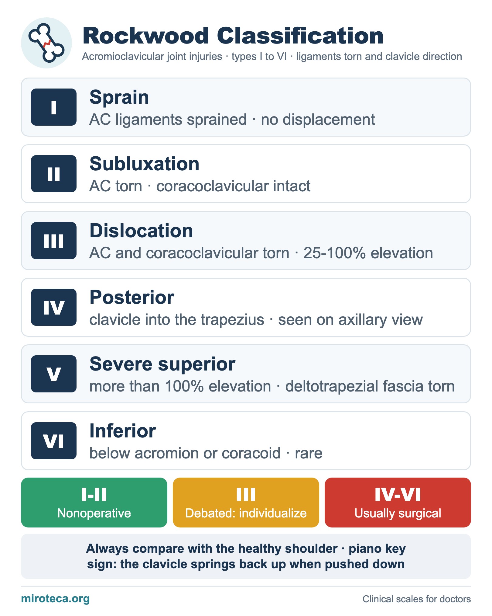
What the chart shows
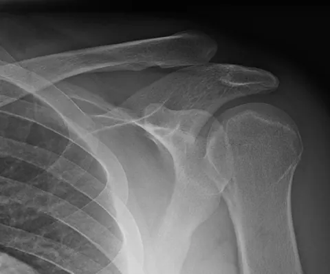
The Rockwood classification grades acromioclavicular (AC) joint injuries, usually from a direct fall on the shoulder, into six types according to the acromioclavicular and coracoclavicular ligaments torn and the direction of clavicle displacement. It expanded the older three-grade Tossy system.
The detail that settles it
Type III tears both ligament groups and raises the clavicle 25-100% above the normal coracoclavicular distance; type V goes beyond 100%. Type IV displaces the clavicle posteriorly into or through the trapezius and is easily missed on the AP view, so an axillary view is needed. Type VI is a rare inferior displacement.
In exam questions
Always compare with the uninjured shoulder. Types I and II are treated nonoperatively, type III is debated and individualized, and types IV to VI are usually surgical.
Keep practicing
This page is the visual summary. To test yourself with real clinical reasoning, Medicinedle, a Wordle-inspired game, gives you one clinical case a day: guess the diagnosis in six tries, with a new clue after every miss. It is free.
Prefer quick questions? The clinical scores quiz gives you 5 case-based questions in about a minute, with the answer explained.